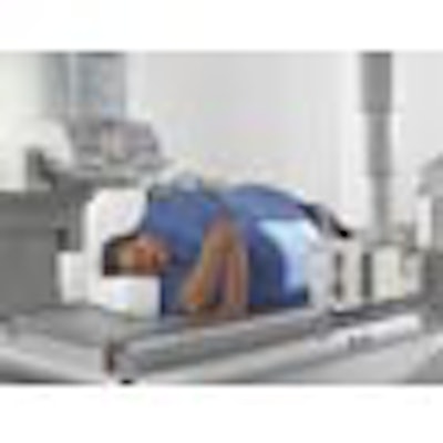
Multimodality vendor Siemens Medical Solutions of Malvern, PA, is introducing a new configuration of its Axiom Artis dBA biplane neurointerventional angiography system, first introduced in 2004.
The older version Axiom Artis dBA paired a large detector with a small detector; the new Axiom Artis dBA twin version has two large detectors. The system can be used for simultaneous digital imaging techniques, and is designed to support modern angiography and interventional procedures in neuroradiology and universal angiography.

Siemens will also be showing enhancements to DynaCT, a rotational angiography upgrade for its Axiom Artis FD C-arm angiography systems that allows soft-tissue imaging in the angiography suite, an application that Siemens is calling angiographic computed tomography (ACT). The technique virtually eliminates the need to transfer patients to other modalities for follow-up procedures. DynaCT has U.S. Food and Drug Administration clearance and is available in the U.S. and worldwide.
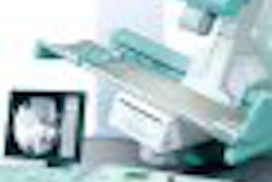
Siemens will demonstrate a compact configuration of the Axiom Aristos FX Plus ceiling-mounted digital angiography system. The system can be moved into position at the touch of a button, enabling lateral and oblique exposures as well as trauma applications. The compact configuration enables customers to install the system in small hospital rooms, according to the company.
Also look for Siemens to highlight Axiom Luminos TF, a conventional fluoroscopy system with a table limit of 600 lb, as well as Axiom Aristos VX Plus, a single-detector radiography system with a 17 x 17-inch flat-panel digital detector.
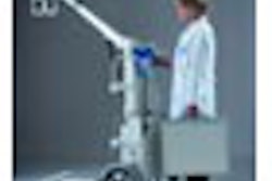
The company's special procedures division will highlight Arcadis Avantic, a mobile C-arm sporting a 13-inch image intensifier, and Arcadis Orbic and Arcadis Orbic 3D, a family of C-arms with an isocentric design and 190° of orbital movement. The company will also tout the Siremobil Compact L and Mobilett XP Digital portable x-ray systems.
By Robert Bruce
AuntMinnie.com contributing writer
October 30, 2006
Copyright © 2006 AuntMinnie.com


![Representative example of a 16-year-old male patient with underlying X-linked adrenoleukodystrophy. (A, B) Paired anteroposterior (AP) chest radiograph and dual-energy x-ray absorptiometry (DXA) report shows lumbar spine (L1 through L4) areal bone mineral density (BMD). The DXA report was reformatted for anonymization and improved readability. The patient had low BMD (Z score ≤ −2.0). (C) Model (chest radiography [CXR]–BMD) output shows the predicted raw BMD and Z score in comparison with the DXA reference standard, together with interpretability analyses using Shapley additive explanations (SHAP) and gradient-weighted class activation maps. The patient was classified as having low BMD, consistent with the reference standard. AM = age-matched, DEXA = dual-energy x-ray absorptiometry, RM2 = room 2, SNUH = Seoul National University Hospital, YA = young adult.](https://img.auntminnie.com/mindful/smg/workspaces/default/uploads/2026/04/ai-children-bone-density.0snnf2EJjr.jpg?auto=format%2Ccompress&dpr=2&fit=crop&h=167&q=70&w=250)


