It sounds like something out of a comic book -- a technique for producing images called dark-field x-ray. But the technology is real, and could offer a way to create higher-quality images using conventional digital radiography equipment.
In a paper published online in Nature Materials on January 20, Swiss researchers at Paul Scherrer Institute and the Ecole Polytechnique Fédérale de Lausanne in Switzerland discussed their implementation of dark-field imaging. The term refers to the creation of x-ray images based on radiation scattered from a target (dark field) rather than the traditional method of using radiation absorbed by the target (light field).
Dark-field imaging isn't a new concept -- it was developed five to seven years ago, according to Franz Pfeiffer, Ph.D., a professor at Paul Scherrer Institute and lead researcher on the Nature Materials study. But previously, dark-field imaging was possible only using x-rays from an extremely brilliant source, such as a synchrotron, a type of particle accelerator that is typically housed in a massive building.
Instead, the Swiss researchers developed a type of grid system designed to measure scatter radiation and create dark-field images with the types of x-ray sources and digital detectors used for conventional medical imaging or nondestructive testing applications. Called an x-ray grating interferometer, one piece of the grid is placed in front of the specimen to "preprint" the x-ray beam; two other grids are placed after the specimen and before the digital detector.
During a study, four images are collected to measure scatter radiation, and the grids are shifted slightly between images. The researchers then examine the resulting image set to detect slight changes created by the specimen on the structure of the x-ray beam.
The images the group has produced so far indicate sharply higher image quality for dark-field x-rays when compared to images produced with conventional absorption-based techniques. Pfeiffer said the difference isn't so much higher spatial resolution for the dark-field technique; rather, certain types of structures -- such as very small cracks or abrupt edges -- produce scatter radiation that shows up well on dark-field images.
Clinical applications
This means that a dark-field x-ray system could be adept at detecting small hairline fractures that might not be visible on a conventional digital x-ray unit. Another promising application could be bone density measurement.
"I'm sure dark-field imaging, in combination with a standard technique, may give you an improved diagnosis," Pfeiffer said. "If you have a complicated fracture in a bone and small splinters, this will show up nicely."
Farther down the road could be an adaptation of dark-field radiography to detect differences in soft tissue of the type that might be present between cancer and normal tissue. Pfeiffer estimates that the technology is one to two years from having the sensitivity required for that application.
Despite its promise, dark-field x-ray could be a ways from your local hospital or imaging center. The closest thing to a human the researchers have studied was a chicken wing purchased from a local grocery store. But the group is in the process of getting regulatory approval for studying human subjects, and Pfeiffer estimates that such trials could be about a year away.
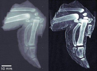 |
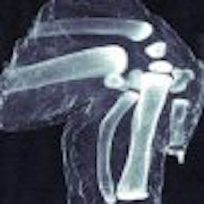
| At left, conventional radiography image of chicken wing. At right, dark-field x-ray image of same chicken wing. Images courtesy of Franz Pfeiffer, Ph.D. |
Assuming that dark-field x-ray does become clinically viable, will it require prospective users to make major modifications to their radiography systems -- or worse yet, purchase an entirely new DR unit? Pfeiffer doesn't think so. Adding a dark-field capability to a DR system would require some changes, but users should be able to use their existing x-ray source and detector assembly.
"To implement the grids you'd need mechanical adaptations, but not new a detector or x-ray source," he said. "It could be an add-on to existing machines."
Pfeiffer said the research group has received some interest from medical imaging vendors as to the commercial potential of a dark-field x-ray system. The introduction of a commercial system "depends on how motivated the manufacturers are," Pfeiffer said. "If you have a lot of money you could get there in three to four years."
By Brian Casey
AuntMinnie.com staff writer
February 4, 2008
Related Reading
New CR technology boosts resolution, bags R&D award, September 3, 2007
Copyright © 2008 AuntMinnie.com
