German researchers have developed a device that could make possible the use of a technique called dark-field CT in clinical imaging of humans, according to a study published February 7 in Proceedings of the National Academy of Sciences.
The dark-field CT technique could provide more useful clinical data because it could measure x-ray properties that conventional CT can't, wrote a team led by Manuel Viermetz of the Technical University Munich in Germany.
"X-ray CT is one of the most commonly used diagnostic three-dimensional imaging modalities today," the group wrote. "Conventionally, this noninvasive technique generates contrast by measuring the x-ray attenuation properties of different tissues. Considering the wave nature of x-rays, complementary contrast can be achieved by further measuring their small-angle scattering (dark-field) properties."
CT shows differences in how x-rays attenuate as they pass through tissues. Some wave properties of x-rays, such as refraction and small-angle scattering, offer useful diagnostic data. But the technology for gathering this data has not been developed to a usable level for medical imaging, the journal noted in a statement.
So Viermetz and colleagues integrated a Talbot-Lau interferometer into a clinical CT gantry to collect tissue microstructure information from small-angle x-ray scattering. The device included data-processing algorithms that mitigated any artifacts caused by the vibration of the CT gantry, as well as tested the system on a phantom of a human thorax.
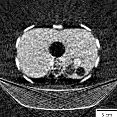
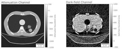
 Dark-field CT reconstruction of a clinical chest phantom. Besides conventional attenuation contrast, dark-field contrast is also retrieved, providing information on the porosity of tissue. Here, the chest phantom is filled with a foam insert that simulates lung tissue, and tubes containing different materials (left-to-right: POM, air, powdered sugar, water). While purpose structures are too small and have low conventional attenuation CT, they are easily visible in the dark-field image. Image and caption courtesy of Manuel Viermetz.
The group found that the device was effective in capturing dark-field data (i.e., small-angle scattering), which "provides additional valuable diagnostic information on otherwise unresolved tissue microstructure," it noted.
"Dark-field computed tomography has been an active research field since more than a decade but was -- until now -- only realized in rather small laboratory setups or at synchrotrons," Viermetz told AuntMinnie.com via email. "In our work, the novelty is the realization of the dark-field technology with a clinical CT [device]."
The research lays a foundation for implementing dark-field CT for real-world medical applications, according to the authors. In fact, once dark-field CT systems were approved for clinical use, the group would expect that they would have "immediate impact" on lung imaging, as the technique has already shown promise in small animal studies on chronic obstructive pulmonary disease, emphysema, fibrosis, and lung cancer.
"The fact that the demonstrated proof of concept requires only minor modifications of a prevalent device allows for rapid translation to clinical application and commercialization," Viermetz and colleagues wrote. "The proposed dark-field CT is not only fully compatible [with existing CT systems] but actually, even advantageous in combination with other innovations (e.g., dual-energy and photon-counting detectors) that are currently introduced by manufacturers."


