An experimental dark-field chest x-ray system can acquire diagnostic attenuation-based x-rays comparable to those from conventional systems, according to a study published February 18 in European Radiology.
A team led by Margarete Kattau of the Technical University of Munich in Germany compared the visual quality of attenuation images obtained with the prototype with those from a commercial radiography system. Overall image quality was rated by readers as high for both devices, a finding that advances the prototype toward clinical use, the researchers suggest.
"Despite a low tube voltage (70 kVp) and comparably long acquisition time, the attenuation images from the dark-field chest radiography system achieved diagnostic quality for lung assessment," Kattau and colleagues wrote.
Dark-field radiography is an imaging approach currently being investigated in clinical trials in patients with chronic obstructive pulmonary disease (COPD). The system is based on modifying existing x-ray scanners with interferometers to visualize the scattering of x-rays in the alveoli of the lungs (the dark-field signal).
Importantly, the prototype enables the acquisition of both dark-field images and attenuation-based images simultaneously, which are then combined in processing to reveal more diagnostic information than either image alone, according to the authors. In this study, the group investigated whether the attenuation-based radiographs alone from the prototype can be used for diagnosis and perhaps eliminate the need for additional conventional x-rays.
Chest images from both the prototype and a conventional commercial system were acquired in 65 patients enrolled in an ongoing clinical trial. In total, 11 patients were categorized as COPD and 51 without COPD.
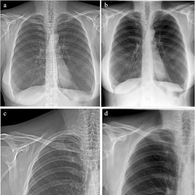
 Attenuation images of a 42-year-old woman with COPD (GOLD III). (A) Attenuation image acquired with the prototype. (B) Attenuation image acquired at the commercial system. Images C and D show enlarged extracts from A and B, respectively. Image courtesy of European Radiology through CC BY 4.0.
Five radiologists independently assessed the visibility of anatomical structures, the level of motion artifacts, and the overall image quality of all attenuation images on a five-point scale, with five points being the highest rating.
Overall image quality was rated high for both devices, 4.2 for the prototype and 4.6 for the commercial system, according to the results. The rating scores varied only slightly between both image types, especially for structures relevant to lung assessment, where the images from the commercial system were graded slightly higher, the authors wrote.
In addition, for the assessment of motion artifacts, the images from the commercial system achieved a mean rating of 4.9, whereas the grating-based images from the prototype scored 4.1 on average.
"Despite the increased presence of motion artifacts and slightly lower average scores in the majority of the criteria, the overall image quality was rated high for the images obtained with our prototype," the authors wrote.
The researchers noted that the prototype requires an acquisition time of seven seconds due to the scanning process. This in turn requires a prolonged breath-hold by patients, which may be a significant limitation for the system in clinical practice, they wrote.
Nonetheless, the results are promising, the group wrote.
"Overall, our results show that the attenuation images acquired with our prototype are of comparable diagnostic quality to the commercial images. This could potentially simplify the implementation of the system into clinical workflow, as it yields both a dark-field and an attenuation image and no additional attenuation x-ray would be required," Kattau and colleagues concluded.