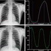
A new study published this month in Radiology by Swedish researchers scores another point for tomosynthesis-based digital radiography (DR) over conventional DR. The study found that tomosynthesis showed better sensitivity in detecting pulmonary nodules, particularly those smaller than 9 mm.
The research team wanted to see if digital tomosynthesis could counteract some of the traditional disadvantages of radiography in chest imaging, such as low sensitivity and specificity. The group was led by Dr. Jenny Vikgren, Ph.D., from Sahlgrenska Academy at the University of Gothenburg in Gothenburg, and included her colleagues from that institution as well as the Sahlgrenska University Hospital, also in Gothenburg. The study was published in Radiology online before print on October 10.
In December 2006, Sahlgrenska University Hospital installed a digital tomosynthesis unit (VolumeRAD option for Definium 8000, GE Healthcare, Chalfont St. Giles, U.K.) and found that the system's moving gantry head offered the potential to visualize pathology obscured by anatomy on conventional 2D radiography. Digital tomosynthesis units collect multiple tomographic slices as they pan across the patient, and these slices can be viewed individually or as part of 3D reconstructions.
Vikgren and colleagues compared digital tomosynthesis to conventional DR (Definium 8000, GE) in a population of 100 patients referred for chest CT between January and May 2007. After the study's exclusion criteria were applied (patients with more than 20 nodules or tomosynthesis images with breathing artifacts), a study sample remained of 42 patients with pulmonary nodules and 47 without. Multidetector CT (MDCT) was used as the reference standard.
For each exam, the digital tomosynthesis unit collected 60 low-dose projection images at a tube voltage of 120 kV over 10 seconds. The effective dose for the patient for the tomosynthesis exam was 0.12 mSv, compared to 0.04 mSv for conventional DR and 4 mSv for multidetector-row CT.
Four thoracic radiologists evaluated the images for the presence of pulmonary nodules, marked the location of suspected lesions, and were asked to grade their confidence that a nodule was present using a score of one to four. The scores were then compared to the number of nodules found on the multidetector CT images.
The results were dramatic: the radiologists found three times as many nodules with tomosynthesis as they did with conventional DR. Tomosynthesis found a total of 121 lesions, or 92% of the 131 nodules found on the reference modality, MDCT. Only 37 lesions (28%) were visible on conventional DR.
In an analysis of each technology's sensitivity based on lesion size, the group found that tomosynthesis performed particularly well in lesions from 4-8 mm, detecting 91% of nodules 4-6 mm and 100% of nodules 6-8 mm. The researchers said that detecting lesions larger than 4 mm is more important in the clinical setting than finding smaller nodules.
Percentage of lesions detected by lesion size
|
The researchers concluded by stating that digital tomosynthesis could be an alternative to conventional radiography, due to its higher sensitivity, as well as to CT, which produces a higher radiation dose and is more expensive. Digital tomosynthesis produced a radiation dose that was 30 times lower than CT, and tomosynthesis dose could be reduced further as dose optimization technologies are developed.
Regarding cost, the researchers noted that the tomosynthesis option added 25% to the cost of a DR unit; this goes up to 40% when additional financial costs are factored in, such as increased image interpretation time and higher archiving costs. Still, the price for a tomosynthesis exam is about 17% of the cost of a routine CT study.
"The detectability of pulmonary nodules is significantly higher for chest tomosynthesis than for chest radiography," the authors wrote. "The sensitivity is increased, especially for nodules smaller than 9 mm. The increased detectability is obtained at a modest increase in radiation dose."
By Brian Casey
AuntMinnie.com staff writer
October 21, 2008
Related Reading
Digital tomosynthesis beats standard DR in finding chest lesions, August 11, 2008
TMJ imaging benefits from digital tomo technique, December 24, 2007
DR pumps new life into tomosynthesis-based radiography, October 29, 2007
Duke researchers combine DR tomosynthesis with 3D CAD, June 4, 2007
Copyright © 2008 AuntMinnie.com


