
Siemens Healthineers has garnered U.S. Food and Drug Administration (FDA) clearance for its Multix Impact floor-mounted digital radiography (DR) system.
Multix Impact features wireless detectors and a free-floating flat tabletop, according to the vendor. In addition, technologists can use a touchscreen user interface on the x-ray tube, as well as continuously monitor the patient from the control room with a patient positioning camera. Graphical program selection and a patient positioning guide are available on both the in-room touchscreen and control room workstation, Siemens said.
Other Multix Impact capabilities include motorization and tracking features designed to reduce physical exertion for staff members, the company said.

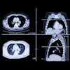
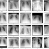


![Representative example of a 16-year-old male patient with underlying X-linked adrenoleukodystrophy. (A, B) Paired anteroposterior (AP) chest radiograph and dual-energy x-ray absorptiometry (DXA) report shows lumbar spine (L1 through L4) areal bone mineral density (BMD). The DXA report was reformatted for anonymization and improved readability. The patient had low BMD (Z score ≤ −2.0). (C) Model (chest radiography [CXR]–BMD) output shows the predicted raw BMD and Z score in comparison with the DXA reference standard, together with interpretability analyses using Shapley additive explanations (SHAP) and gradient-weighted class activation maps. The patient was classified as having low BMD, consistent with the reference standard. AM = age-matched, DEXA = dual-energy x-ray absorptiometry, RM2 = room 2, SNUH = Seoul National University Hospital, YA = young adult.](https://img.auntminnie.com/mindful/smg/workspaces/default/uploads/2026/04/ai-children-bone-density.0snnf2EJjr.jpg?auto=format%2Ccompress&dpr=2&fit=crop&h=167&q=70&w=250)


