Chest dynamic digital radiography (DDR) may have received a boost toward clinical use in patients with lung disorders, with researchers developing AI to perform time-consuming analysis involved in the technology, according to researchers in New York City.
A group at Mount Sinai Hospital developed a “pipeline” of convolutional neural networks (CNNs) to analyze lung areas in DDR image sequences from patients. The model performed well enough to act as a surrogate to standard pulmonary function tests, they found.
“Our findings add to growing evidence suggesting DDR as a potential [pulmonary function test] surrogate,” noted lead author and internal medicine fellow Valeria Santibanez, MD, and colleagues. The study was published March 29 in Chest Pulmonary.
Common diagnostic tests for pulmonary disorders include chest x-rays and pulmonary function tests (PFTs). Although essential, these tests offer a limited static assessment, the authors wrote. Alternatively, previous research has shown that chest DDR can capture active lung movement during respiration by offering a detailed view of lung and diaphragm motion.
DDR is relatively easy to obtain, but barriers to its clinical adoption include time-consuming manual analysis and unclear correlation with standard PFTs, the authors noted.
To that end, the group developed two convolutional neural networks (CNN) designed to quantify key measurements in DDR image sequences. For data, they used PFT and DDR studies from 55 patients with normal, obstructive, and restrictive lung physiology.
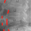
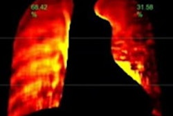
 (a) Raw example of a dynamic digital radiograph. (b) Contrast equalization using contrast-limited adaptive equalization shows improved anatomical visualization. (c) Training data is generated through manually labeling using SLEAP’s graphical user interface. (d-e) Neural network detection schematic. For each quadrant a series of two networks were used. The first identifies the quadrant center, the second using input of the first, then identifies individual anatomical landmarks within a quadrant. (d) Four neural networks were trained, each with the objective of identifying a chest quadrant centroid. The four separate quadrants are visualized as colored boxes (right upper quadrant, green; right lower quadrant, blue; left upper quadrant, red; left lower quadrant, yellow). (e) Serial centroid then anatomical identification. Two quadrant examples showing confidence maps of trained neural networks identifying quadrant centroids (left) and then anatomical confidence maps (right). Image and caption courtesy of Chest Pulmonary.
The DDR sequences were acquired using commercially available DDR machines (Konica Minolta Healthcare Americas, Wayne, NJ) and the CNNs were trained to quantify lung areas and generate what the group termed “DDR-based PFT’s,” or dPFTs. Next, the team compared specific PFT and dPFT measurements.
According to the analysis, the researchers observed statistically significant, strong correlations between dPFT areas and standard PFT volumes, including total lung capacity (r = 0.764), forced expiratory volume in one second (r = 0.591), vital capacity (r = 0.763), and functional residual capacity (r = 0.756).
“Statistically significant correlations found between dPFTs and PFTs suggest that dPFTs can act as a surrogate to PFTs when these are not available or unable to be performed,” the group wrote.
Ultimately, DDR has unique advantages over traditional studies, the authors noted. The digital technology limits radiation exposure to patients compared with standard chest x-rays, they wrote. Moreover, while PFTs are vital for diagnosing and monitoring pulmonary disorders, they pose challenges in accessibility, such as in patients with neuromuscular conditions or those experiencing flares of chronic obstructive pulmonary disease, they suggested.
“Our study demonstrates robust dPFTs and PFTs correlations using an automated DDR analysis pipeline. This pipeline has potential to discern normal from abnormal physiology, suggesting dPFTs are valuable in assessing lung dynamics,” the group concluded.
The full article is available here.