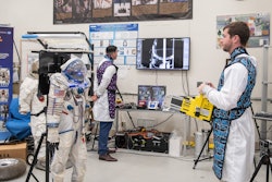
In the next few days, astronauts aboard a SpaceX mission will take x-rays for the first time in space.
The Fram2 mission launched on March 31 at 9:46 p.m. EDT from NASA's Kennedy Space Center in Florida. The crew will be in orbit for three to five days and will conduct more than 20 science experiments, including taking images of each other with an ultraportable x-ray machine.
In an interview with AuntMinnie.com, radiologist Michael Pohlen, MD, of the University of California, San Diego, and a consultant on the mission, said that this first use of x-ray in orbit is mostly a technical demonstration.
“It's mostly to prove that the equipment works and we can acquire images that are diagnostic and that this tool can then be used in the future to assist in more hypothesis-driven work,” he said.
Of major concern is that astronauts have been shown to lose about 1% of their bone mineral density per month in space due to the lack of gravitational force on bones. The use of portable x-ray could be a way to measure these changes “in flight,” Pohlen noted.
Pohlen, who penned a 2023 essay in the American Journal of Roentgenology about his early days working at NASA, said that the x-ray experiments have heralded in a new era for radiology in space medicine, with all previous imaging research involving the use of ultrasound.
In continuing coverage, AuntMinnie.com will be featuring interviews with the industry partners on the experiments, the x-ray detector’s developer, KA Imaging, and the maker of the device itself, MinXray.
![Representative example of a 16-year-old male patient with underlying X-linked adrenoleukodystrophy. (A, B) Paired anteroposterior (AP) chest radiograph and dual-energy x-ray absorptiometry (DXA) report shows lumbar spine (L1 through L4) areal bone mineral density (BMD). The DXA report was reformatted for anonymization and improved readability. The patient had low BMD (Z score ≤ −2.0). (C) Model (chest radiography [CXR]–BMD) output shows the predicted raw BMD and Z score in comparison with the DXA reference standard, together with interpretability analyses using Shapley additive explanations (SHAP) and gradient-weighted class activation maps. The patient was classified as having low BMD, consistent with the reference standard. AM = age-matched, DEXA = dual-energy x-ray absorptiometry, RM2 = room 2, SNUH = Seoul National University Hospital, YA = young adult.](https://img.auntminnie.com/mindful/smg/workspaces/default/uploads/2026/04/ai-children-bone-density.0snnf2EJjr.jpg?auto=format%2Ccompress&fit=crop&h=100&q=70&w=100)


![Representative example of a 16-year-old male patient with underlying X-linked adrenoleukodystrophy. (A, B) Paired anteroposterior (AP) chest radiograph and dual-energy x-ray absorptiometry (DXA) report shows lumbar spine (L1 through L4) areal bone mineral density (BMD). The DXA report was reformatted for anonymization and improved readability. The patient had low BMD (Z score ≤ −2.0). (C) Model (chest radiography [CXR]–BMD) output shows the predicted raw BMD and Z score in comparison with the DXA reference standard, together with interpretability analyses using Shapley additive explanations (SHAP) and gradient-weighted class activation maps. The patient was classified as having low BMD, consistent with the reference standard. AM = age-matched, DEXA = dual-energy x-ray absorptiometry, RM2 = room 2, SNUH = Seoul National University Hospital, YA = young adult.](https://img.auntminnie.com/mindful/smg/workspaces/default/uploads/2026/04/ai-children-bone-density.0snnf2EJjr.jpg?auto=format%2Ccompress&fit=crop&h=112&q=70&w=112)


