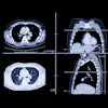
The Fram2 mission splashed down safely on April 4 at 9:19 a.m. PT off the coast of Oceanside, CA, counting among its accomplishments the first use of an x-ray machine in space.
While the mission provided a striking in-flight image, the spectral imaging capabilities of the ultraportable x-ray machine’s detector offer researchers a much deeper dive into human anatomy, according to Karim Karim, PhD, chief technology officer of KA Imaging.
“Because of spectral technology for the first time you can do quantitative imaging with x-ray,” Karim said.
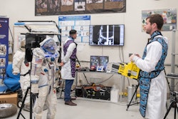
The Fram2 mission launched on March 31, and special considerations included how vibrations during liftoff could affect the x-ray device, as well as how increased space radiation could affect its performance, Karim noted.
KA Imaging, based in Waterloo, Ontario, received U.S. Food and Drug Administration clearance for the detector in 2020, and aside from space, the technology is also revolutionizing imaging in remote and underserved areas on Earth, Karim said.
In part I of the series, we interviewed radiologist Michael Pohlen, MD, a consultant on the Fram2 mission. Stay tuned for part III, an interview with MinXRay, the portable x-ray machine’s developer.


![Representative example of a 16-year-old male patient with underlying X-linked adrenoleukodystrophy. (A, B) Paired anteroposterior (AP) chest radiograph and dual-energy x-ray absorptiometry (DXA) report shows lumbar spine (L1 through L4) areal bone mineral density (BMD). The DXA report was reformatted for anonymization and improved readability. The patient had low BMD (Z score ≤ −2.0). (C) Model (chest radiography [CXR]–BMD) output shows the predicted raw BMD and Z score in comparison with the DXA reference standard, together with interpretability analyses using Shapley additive explanations (SHAP) and gradient-weighted class activation maps. The patient was classified as having low BMD, consistent with the reference standard. AM = age-matched, DEXA = dual-energy x-ray absorptiometry, RM2 = room 2, SNUH = Seoul National University Hospital, YA = young adult.](https://img.auntminnie.com/mindful/smg/workspaces/default/uploads/2026/04/ai-children-bone-density.0snnf2EJjr.jpg?auto=format%2Ccompress&dpr=2&fit=crop&h=167&q=70&w=250)


