An AI model appears to be a practical and reliable tool for predicting osteoporosis from frontal lumbar spine x-rays, researchers in Japan have reported.
The finding is from a study involving 332 patient x-rays from which the model estimated bone mineral density, with its estimations correlating strongly with gold standard dual-energy x-ray absorptiometry (DEXA) scans, noted lead author Ryusei Inamori, a doctoral student at Tohoku University in Miyagi, Japan, and colleagues.
“[The software] may provide screening opportunities for patients in various medical settings and contribute to extending healthy life expectancy among the elderly through early detection,” the group wrote. The study was published in the January-March issue of the Journal of Clinical Densitometry.
Osteoporosis affects approximately 200 million people worldwide. DEXA scanners are available only in limited facilities, which can hinder early screening for the disease, the authors explained.
To date, several AI models have been developed that predict osteoporosis by estimating bone mineral density (BMD) from x-rays, yet these models have generally used data from geographically homogeneous populations, the group noted. One such model (DeepXray Spina, Alpha Intelligence Manifolds) was developed using approximately 15,000 DEXA scans from four medical institutions in Taiwan. For this study, the researchers tested its generalizability across a Japanese cohort.
The group gathered data from 328 patients who underwent both lumbar spine x-rays and DEXA scans within 30 days at Tohoku University Hospital between May 2014 and April 2024. Patients ranged in age from 13 to 91 years old, with 126 classified as normal, 118 as having osteopenia, and 84 as having osteoporosis. The researchers compared assessments based on DEXA and estimates from the AI model, which detects key anatomical landmarks from the T12 to L5 vertebrae and then outputs estimated BMD using machine learning.
According to the analysis, the correlation between the AI-estimated and observed DEXA BMD was strong, with a Pearson’s correlation coefficient of 0.901 and a normalized root mean square error of 0.070, the researchers reported.
Osteoporosis classification from spinal x-rays using an AI model | |||
Measure | Normal | Osteopenia | Osteoporosis |
| Accuracy | 90.2% | 100% | 84.2% |
| Sensitivity | 85.4% | 72.9% | 92.4% |
| Specificity | 95.1% | 81% | 100% |
“This study demonstrated that the software achieved high predictive performance in the Japanese cohort,” the group wrote.
The fact that the AI model requires only lumbar spine x-rays is an advantage, it noted, as x-rays are widely implemented and highly accessible. Thus, the software could provide screening opportunities for patients who visit hospitals for purposes unrelated to osteoporosis (opportunistic screening), the team added.
Also, x-rays can be performed using portable devices, which means that the software could provide screening opportunities to patients who may have difficulty accessing medical facilities, the researchers wrote.
“Thus, the software is a practical tool for predicting osteoporosis and may provide screening opportunities for patients in various medical settings,” they concluded.
The full study is available here.


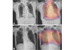
![Representative example of a 16-year-old male patient with underlying X-linked adrenoleukodystrophy. (A, B) Paired anteroposterior (AP) chest radiograph and dual-energy x-ray absorptiometry (DXA) report shows lumbar spine (L1 through L4) areal bone mineral density (BMD). The DXA report was reformatted for anonymization and improved readability. The patient had low BMD (Z score ≤ −2.0). (C) Model (chest radiography [CXR]–BMD) output shows the predicted raw BMD and Z score in comparison with the DXA reference standard, together with interpretability analyses using Shapley additive explanations (SHAP) and gradient-weighted class activation maps. The patient was classified as having low BMD, consistent with the reference standard. AM = age-matched, DEXA = dual-energy x-ray absorptiometry, RM2 = room 2, SNUH = Seoul National University Hospital, YA = young adult.](https://img.auntminnie.com/mindful/smg/workspaces/default/uploads/2026/04/ai-children-bone-density.0snnf2EJjr.jpg?auto=format%2Ccompress&fit=crop&h=167&q=70&w=250)