Structured reporting with AI assistance improves efficiency and diagnostic accuracy when radiologists report on bedside chest x-rays, according to a study published February 3 in Radiology.
The finding is from a comparative reader study that evaluated the effects of three reporting modes on the diagnostic workflow: free-text reporting, structured reporting, and structured reporting with AI-prefilled suggestions, noted lead author Mahta Khoobi, of University Hospital Aachen in Germany, and colleagues.
“Structured reporting of chest radiographs improved diagnostic efficiency, streamlined visual search patterns, shifted focus from the report to the radiograph, and, when supplemented with AI-based suggestions, enhanced diagnostic accuracy,” the group wrote.
Given the growing demand for radiology services and increasing imaging volumes and complexity, structured reporting and AI tools have the potential to transform workflows, the authors noted. While it is critical to develop reporting workflows that balance efficiency and accuracy while minimizing distractions, little is known about how different reporting modes -- and AI assistance -- impact diagnostic workflows, they added.
To bridge the gap, the group conducted a prospective study between July and December 2024 in which they recruited four novice readers and four non-novice readers who each analyzed 35 bedside chest x-rays using the three reporting modes. The AI predictions were generated using a standard convolutional neural network that the researchers previously trained and validated using a dataset of 122,294 chest x-rays.
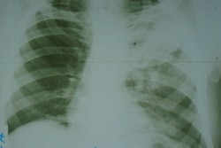
Clinical indications for the images in the study included evaluation of cardiopulmonary status, guidance of fluid management, and assessment of pleural effusions, with the ground truth established by six expert radiologists.
The researchers set up a customized viewer to display the x-rays and the reporting interface on a screen-based eye-tracking system. Outcome measures included diagnostic accuracy, reporting time per radiograph, and eye-tracking metrics.
Key findings included the following:
AI-prefilled structured reporting (AI-SR) improved diagnostic accuracy for all readers (kappa = 0.71; p < 0.001), with both novice readers and non-novice readers showing improvement with AI-SR compared with free-text reporting (delta kappa = 0.17 and 0.09; both p < 0.001).
Radiograph reporting time decreased from 88.1 seconds with free-text reporting to 37.3 seconds with structured reporting and further to 25 seconds with AI-SR (p < 0.001 for all).
Structured reporting shifted readers’ visual attention from the report to the x-ray: Mean total report fixation duration was 11.4 seconds for free-text reporting, 4.8 seconds for structured reporting, and 3.6 seconds for AI-SR (p < 0.001).
 Heatmap overlays of fixations on the viewer display as a function of reader experience level and reporting mode. Each panel shows combined fixation heatmaps for all readers in each group. Within each panel, the radiograph display field is located on the right, and the corresponding report display field is on the left. The heatmaps illustrate the spatial distribution of cumulative fixation durations, with green indicating lower fixation intensity and red indicating higher fixation intensity. Color scaling was applied individually to each heatmap, meaning that fixation intensities are not directly comparable across panels. Heatmaps were generated in the postprocessing stage using Tobii Studio. With structured reporting and AI-prefilled structured reporting, readers’ visual attention was particularly focused on the key anatomic regions queried by the structured template. As a result, gaze was centered on the central and basal lung fields, whereas the lung apices and extrapulmonary or extracardiac structures received less attention.RSNA
“Reporting mode significantly impacted diagnostic accuracy and efficiency in this study. Structured reporting enhanced efficiency by directing visual attention toward the image, particularly benefiting inexperienced readers. Artificial intelligence (AI)-prefilled SR improved diagnostic accuracy,” the group wrote.
Future studies should use eye trackers that integrate into the workflow, include actual radiology workstations with multiple screens, provide access to prior imaging studies and clinical information, and allow image manipulation functions such as zooming and panning, the researchers noted.
“They should also incorporate bias-control strategies, emphasize transparency and explainability, and confirm robustness through multicenter evaluation and prospective trials before clinical adoption,” the group concluded.
The full study is available here.