A study from the Cleveland Clinic to investigate brain metabolism of patients with amyotrophic lateral sclerosis (ALS, or Lou Gehrig's disease) has shown that PET imaging may be useful in distinguishing typical dementia syndromes, such as Alzheimer's disease and frontotemporal dementia, from atypical cortical changes.
In addition, PET scans demonstrated that the ALS patients with cognitive impairment represent a heterogeneous group.
Although they may have specific dementia syndrome, such as Alzheimer's disease, frontotemporal dementia, or aphasia syndrome, the majority of patients are either normal or show diffuse cortical hypometabolism.
Early diagnosis
ALS is a rare, neurodegenerative disorder that affects mainly the motor neurons and ultimately leads to death. "Earlier diagnosis might lead to earlier treatment and earlier involvement in clinical trials, which also could help monitor the disease process," said Dr. Roland Talanow, study co-author and presenter at the RSNA conference.
The study included 11 patients (eight females and three males) with a mean age of 65 years. Every patient was clinically diagnosed with ALS and underwent PET imaging for signs of cognitive impairment. Imaging was performed on a Siemens Healthcare ECAT HR+ PET scanner. Patients were injected intravenously with FDG at a dose of 5 to 15 mCi, followed by an uptake time of 35 to 40 minutes.
Researchers collected the PET data over a 26-month period. A neurologist who specializes in ALS evaluated the information.
The researchers measured the level of PET metabolism and divided the results into three categories -- normal metabolism, hypometabolism, and hypermetabolism. The normal metabolism group also was divided into frontal, temporal, parietal, occipital, and diffuse segments. Regions of abnormal metabolism were categorized by right and left cerebral hemisphere.
Patient results
In the analysis, all patients demonstrated a certain degree of hypometabolism in the primary motor cortex as expected for ALS. "In the other regions of the cortex," Talanow added, "three patients appeared normal for their age and eight patients demonstrated regional hypometabolism in the nonmotor cortex areas."
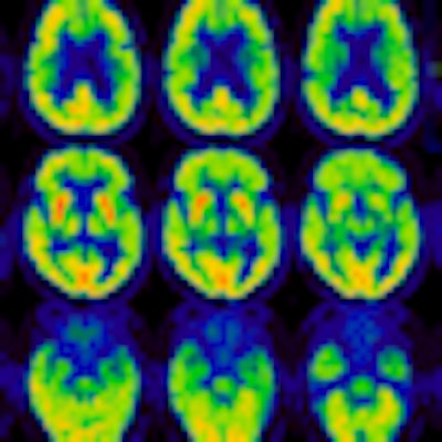
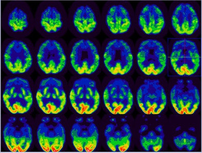
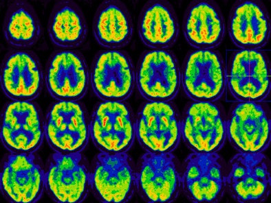
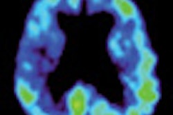
Of the eight patients with abnormal cortical hypometabolism, two patients demonstrated a pattern of frontotemporal dementia, while one patient was consistent with Alzheimer's disease. One patient demonstrated asymmetric left frontal, parietal, temporal hypometabolism reminiscent of progressive aphasia syndrome. The remaining four patients demonstrated diffuse cortical hypometabolism.
 |
| In this image of frontotemporal dementia, there is severely reduced FDG activity in the bilateral anterior temporal, bilateral frontal, and bilateral anterior parietal regions. Mild to moderate to decreased FDG uptake is most noted in mid to posterior temporal and regions. This finding is consistent with frontotemporal dementia. All images courtesy of Dr. Roland Talanow and the Cleveland Clinic. |
 |
| In this image of diffuse cortical hypometabolism, there is moderately reduced FDG activity in the entire cerebral cortex relative to the basal ganglia. The pattern of diffuse hypometabolism is not suggestive of specific dementia syndrome. The finding is more compatible with diffuse metabolic insult or medication effect. |
In conclusion, the study found that ALS patients with cognitive impairment are a heterogeneous group on PET imaging. "Although ALS may manifest as specific dementia syndrome, such as frontotemporal dementia or Alzheimer's dementia, the majority of patients expressed nonspecific focal or diffuse cortical hypometabolism," Talanow said.
While the utility of PET is not yet fully established in ALS, the authors opined that PET is "likely" a good resource to further characterize cortical dysfunction of ALS patients.
PET also may be a "useful tool separating typical dementia syndrome from atypical cortical changes," the researchers wrote.
By Wayne Forrest
AuntMinnie.com staff writer
December 2, 2008
Related Reading
PET, PiB support cognitive reserve hypothesis in Alzheimer's, November 14, 2008
WSJ: FDA supports imaging agents for Alzheimer's, October 24, 2008
PET with C-11 PiB can assess beta-amyloid brain deposits, August 12, 2008
PET scans help spot and classify dementia types, March 27, 2008
FDG-PET CAD scheme boosts diagnostic accuracy for Alzheimer's, October 2, 2007
Copyright © 2008 AuntMinnie.com


