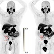
Korean researchers have developed a new PET imaging radiotracer that appears equal to gallium-68 (Ga-68) prostate-specific membrane antigen (PSMA)-11 for detecting primary and metastatic lesions in patients with prostate cancer, according to a study in the October 1 issue of the Journal of Nuclear Medicine.
In a head-to-head comparison, a team at Seoul National University pitted a radiotracer they created called Ga-68 NOTA Glu-Urea-Lys (NGUL) against Ga-68 PSMA-11 to compare the detection efficacy and biodistribution between the two tracers in the same patients with metastatic prostate cancer.
"The ability to detect primary and metastatic lesions was identical between Ga-68 NGUL and Ga-68 PSMA-11," wrote a team led by professor of nuclear medicine Dr. Gi Jeong Cheon, PhD.
PSMA is a transmembrane protein overexpressed in prostate cancer, the most common cancer in men. The overexpression of PSMA is directly related to tumor grade, cancer progression, and the presence of metastases.
PET radiotracers have been designed to attach to the PSMA protein. The radiotracers emit positron particles detectible with PET imaging, and thus highlight the primary location of the tumor as well as areas where the cancer has spread.
In December 2020, Ga-68 PSMA-11 became the first PET radiotracer approved by the U.S. Food and Drug Administration for targeting lesions positive for prostate-specific membrane antigen. Dozens of other radiotracers are currently in development.
Cheon and colleagues synthesized and tested Ga-68 NGUL in mice in 2018 and found it showed a higher tumor-to-background ratio and substantially lower kidney uptake than Ga-68 PSMA-11. In this study, the researchers sought to further investigate the clinical feasibility of Ga-68 NGUL.
The researchers prospectively recruited 11 patients with metastatic prostate cancer. Each patient underwent both Ga-68 NGUL and Ga-68 PSMA-11 PET/CT between one and four days apart, with no patient receiving any treatment between the scans. Scans were obtained at 60 minutes after tracer injection.
Quantitative tracer uptake was determined in normal organs and in primary and metastatic lesions. Lesion numbers and lesion uptake were measured by SUVmax and then compared.
In a total, the researchers identified 161 nodal and 59 bone PSMA-avid metastases in the 11 patients. All lesions were detected identically by both tracers, and none of the lesions were detected by only Ga-68 NGUL or only Ga-68 PSMA-11, they found.
In addition, the researchers evaluated quantitative uptake in a total of 36 lesions (20 lymph nodes and 16 bone metastases). They selected a total of five lesions in each patient. Again, no significant differences in lymph node or bone metastasis uptake were observed between the two tracers, the authors found.
"There was no significant difference in absolute lesion uptake; however, tumor-to-background ratio tended to be lower for Ga-68 NGUL than for Ga-68 PSMA-11," the team stated.
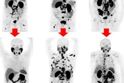
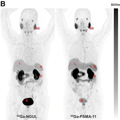
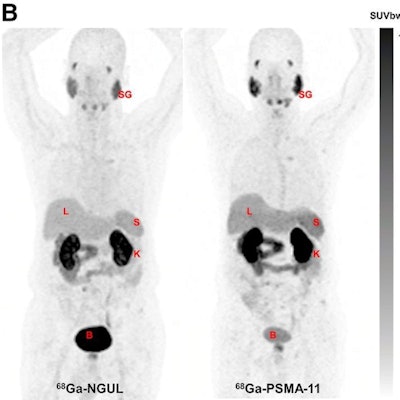 Representative image showing normal-organ distribution of Ga-68 PSMA-11 and Ga-68 NGUL. B = bladder; K = kidney; L = liver; S = spleen; SG = salivary glands; SUVbw = SUV body weight. Image courtesy of the Journal of Nuclear Medicine.
The authors noted the study was limited by a small number of patients, and so stopped short of generalized conclusions. However, given this was a head-to-head comparison and that differences between the radiotracers were insignificant, the authors concluded that Ga-68 NGUL could be a valuable option for PSMA-PET/CT imaging.
"Nonetheless, further studies with a larger number of patients are needed to validate our findings," the researchers wrote.


