Hybrid PET/MRI imaging with a new radiotracer based on copper-64 can identify blood clots in patients with atrial fibrillation (AFib), according to a study published October 13 in JACC: Cardiovascular Imaging.
A team led by cardiologists at Harvard Medical School performed hybrid PET/MRI scans in patients diagnosed with atrial fibrillation (irregular heart rhythm) using an experimental radiotracer (Cu-64 FBP8) that binds to fibrin, a protein involved in forming blood clots. They found the method performed as well as current standard diagnostic approaches.
"PET/CMR of the fibrin-binding PET probe Cu-64 FBP8 can detect acute to subacute [left atrial appendage] thrombi with a sensitivity of 100% and specificity of 84%, matching the performance of current more invasive approaches," wrote first author David Izquierdo-Garcia, PhD, of Harvard and the Massachusetts Institute of Technology, and colleagues.
The finding suggests PET imaging with Cu-64 FBP8 could be valuable for revealing the source of embolism in patients with transient ischemic attacks and stroke, the authors wrote.
The detection of blood clots in the left atrial appendage (LAA), a small sac in the top left chamber of the heart, is vital in the prevention of stroke. Current methods for identifying such blood clots include transesophageal echocardiography, CT, and cardiac MRI. Although these approaches are of major clinical use, they frequently employ contrast agents, have limited anatomical coverage, and involve heavy sedation, according to the authors.
After previously developing Cu-64 FBP8 in lab studies, the researchers performed studies in rats in 2015 that established the tracer's potential for imaging deep vein thrombosis. In this study, they tested Cu-64 FBP8 for detecting blood clots in the LAA for the first time in humans.
The researchers enrolled 24 patients who presented for treatment for atrial fibrillation at Massachusetts General Hospital. The patients fell into three groups after LAA ultrasound exams: those with no sign of blood clots (n = 12), those with spontaneous blood clots (n = 4), and patients in whom blood clots in the LAA were purposefully induced through the placement of a closure device (n = 8).
Closure devices are implants designed to reduce the risk of strokes that originate in the LAA. They block blood clots that form in the LAA from entering the bloodstream, where they can lead to strokes. All patients underwent PET/cardiac MRI with Cu-64 FBP8 within 14 days of their ultrasound exams.
The researchers found PET imaging with Cu-64 FBP8 detected all of the blood clots identified in patients on ultrasound. In contrast, they observed no evidence of focal Cu-64 FBP8 uptake in patients with negative ultrasound results.
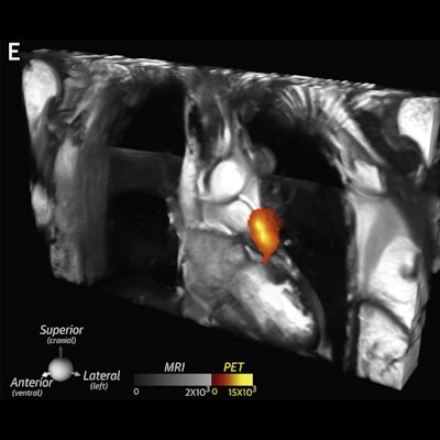
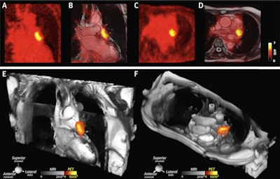 A large thrombus is seen in the left atrial appendage (LAA) behind the device on (A and B) the coronal PET and PET/cardiac MRI images as well as (C and D) the axial PET and PET/cardiac MRI images. (E and F) 2D balanced steady state free precession cines stacks in the coronal and axial planes have been combined into a single 3D dataset and fused with the 3D PET data. In these multimodality images, the PET and MR data are both displayed in 3D, creating a volumetric depiction of the heart and the thrombus/Cu-64 FBP8 containing LAA. Volume-rendered images in the oblique coronal and axial planes confirm the presence of thrombus in the LAA. (A to F) The thrombus produced by the closure device is contained within the LAA, and no evidence of thrombus is seen elsewhere in the heart or thorax. Image courtesy of JACC: Cardiovascular Imaging.
SUVmax values in the LAA were significantly higher in patients with clots in the LAA than those without. An SUVmax threshold of 2.6 yielded a sensitivity of 100% and specificity of 84%, as evaluated by a radiologist with 15 years of experience, the researchers wrote.
In addition, the blood clots induced by the placement of the occlusion device were extremely conspicuous, the authors wrote. All blood clots in these patients were confined to the LAA, behind the device, and they observed no evidence of communication between the LAA and left atrium on images.
"[Cu-64 FBP8-PET/MRI] allows the composite burden of arterial thrombosis and venous thrombi, particularly those in larger veins with the potential to embolize, to be imaged," the researchers wrote.
Furthermore, an analysis of cardiac MRI with T1 mapping in patients with positive ultrasound exams revealed the presence of a hyperintense signal consistent with LAA blood clots, the researchers added. The integrated PET/MRI approach provided useful information on the biological properties of blood clots, such as fibrin and methemoglobin, a byproduct of clots.
Ultimately, the number of spontaneous LAA blood clots identified in patients in the study was low, but they were sufficient to demonstrate proof-of-principle in this first-in-human study, the researchers wrote.
"Further study will be needed to determine the sensitivity of Cu-64 FBP8 for chronic thrombi (> 10 weeks), the utility of adding a gadolinium-based contrast agent to facilitate their detection, and the embolic risk of these fibrin-poor organized thrombi," the authors concluded.


