PET imaging with a gallium-68 (Ga-68)-based radiotracer is more precise than standard MRI for detecting meningiomas targeted for radiotherapy, according to a study published April 20 in the International Journal of Radiation, Oncology, Biology, Physics.
A U.S. group compared Ga-68 DOTATATE PET/CT and contrast-enhanced MRI approaches for treatment planning in patients with meningiomas. They found that clinicians identified additional areas of disease on PET compared with MRI alone.
"Ga-68 DOTATATE PET has great potential to improve local control and progression-free survival while reducing treatment toxicity for patients," wrote Dr. Haley Perlow, a radiation oncologist at the University of Ohio in Columbus.
Meningiomas are tumors that form on the membranes that cover the brain and spinal cord just inside the skull. They have an estimated incidence of 30,000 cases per year in the U.S. Although most meningiomas are benign (grade 1), atypical (grade 2), and malignant (grade 3), subtypes are more aggressive and are often more difficult to detect.
The current standard of care for meningiomas includes MR-based imaging to define the tumors, followed by radiation therapy. PET imaging with Ga-68 DOTATATE, a radiotracer that binds to meningioma cancer cells, has shown better performance than MRI at defining gross tumor volume, but the practice has not been widely adopted.
Perlow and colleagues, who included researchers at the Medical College of Wisconsin in Milwaukee, retrospectively included 25 patients with newly diagnosed or recurrent meningioma. Patients received both Ga-68 DOTATATE PET and contrast-enhanced MRI scans for radiation treatment planning between 2018 and 2020.
Four radiation oncologists and three neuroradiologists were instructed to contour visible tumors on both scans to create gross tumor volumes, which were then compared. Gross tumor volumes have important implications when clinicians determine radiotherapy doses, since controlling tumors requires higher doses if the tumor cell numbers are larger.
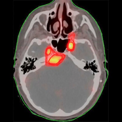
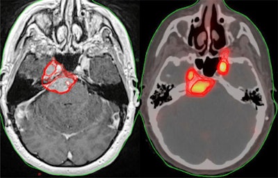
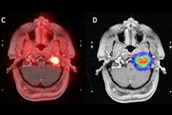
 Patient Z is a female with a history of a grade 1 petroclival meningioma who was found to have progression on her most recent MRI brain scan with an increase in size from 3 x 1.5 cm to 3.4 x 1.4 cm. There was separate uptake on PET within the region of the left temporal lobe, which could relate to extension of the primary mass or separate focus of tumor. She was planned for 54 Gy of radiation in 30 fractions. Image courtesy of the International Journal of Radiation, Oncology, Biology, Physics.
Overall, 344 contoured gross tumor volumes were included in the analysis. The median MRI volume for each physician ranged from 16.94 to 25.53 ccs. Conversely, the median PET volumes for each physician ranged from 2.09 to 8.36 ccs. The median PET volume was smaller for each physician, the researchers found.
In addition, seven of the 25 patients (28%) had new nonadjacent tumor areas contoured on PET by at least six of the seven physicians that were not contoured by these physicians on the corresponding MRI. In other words, these new tumor areas would not have been included in the standard MRI-based volumes, the researchers stated.
"Our study supports that Ga-68 DOTATATE PET imaging may help radiation oncologists create more precise radiation treatment volumes through finding undetected areas of disease not seen on MRI," the authors wrote.
Ultimately, the upshot of the study is that seven physicians were able to independently draw more conformal meningioma tumor volumes on PET scans versus MRI, which provides evidence that Ga-68 DOTATATE PET use can lead to consistent conclusions among different physicians, the authors wrote.
"Ga-68 DOTATATE PET-guided radiation treatments are worthy of future prospective investigation," Perlow and colleagues concluded.


