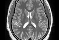
F-18 FDG-PET for the first time has revealed early morphological brain abnormalities that weren't detected by MRI in children with cerebral palsy, according to a group of nuclear medicine researchers in Shiyan, China.
A team at Hubei University of Medicine investigated F-18 FDG patterns of cerebral metabolism changes in children diagnosed with cerebral palsy who showed no structural abnormalities on MRI. The group found distinctive patterns in several brain regions, and it suggests that the findings could help in efforts to diagnose the disorder earlier.
"It is important to diagnose patients with [cerebral palsy] as soon as possible to enable early interventions and accurate evaluation of any therapeutic effects," wrote corresponding author Dr. Zhijun Pei, PhD, and colleagues (September 15, Frontiers in Neurology).
Cerebral palsy is a group of disorders that affect movement and muscle tone or posture. It is caused by damage that occurs to the immature, developing brain, most often before birth. These brain structure abnormalities can be detected using MRI, yet about 15% of children show no abnormalities in their MRI scans, according to the authors.
PET has recently been introduced for monitoring gene therapy for cerebral palsy patients, which suggests it also may be a helpful approach for diagnosing the disorder, according to the authors. Thus, the group explored whether there are correlations between motor function in patients and F-18 FDG brain glucose metabolism.
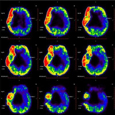
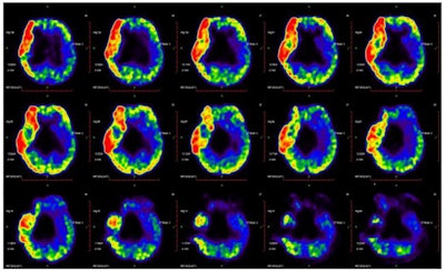
 Pattern related to cerebral palsy is identified by analysis of F-18 FDG-PET scans from 31 patients. The glucose metabolism in the left cerebellum is relatively higher than in the right hemisphere. Image courtesy of Frontiers in Neurology.
The researchers culled imaging data from 31 cerebral palsy patients between the ages of 2 and 4 who had undergone MRI scans at Taihe Hospital in Shiyan, China, that showed no structural brain abnormalities. During clinical evaluation, the motor, linguistic, and cognitive functions of patients were assessed using the Gross Motor Function Classification System (GMFCS), a clinical tool used to classify the severity of motor function deficits.
Images of F-18 FDG cerebral glucose metabolism were acquired (Biograph mCT 64, Siemens Healthineers) with children placed supine on the scan table after sedation. The scans lasted for 45 minutes. Concentrations of F-18 FDG radiotracer were calculated using mean standardized uptake value (SUVmean), with results visually evaluated by two experienced nuclear medicine physicians.
In the individual analysis, most patients showed left > right asymmetry of glucose metabolism in the central region, cerebellum, frontal lobe, and parietal lobe. Glucose metabolism in these left regions was significantly higher than those of the corresponding regions in the right hemisphere, according to the findings.
In addition, levels of motor function measured by the GMFCS were significantly negatively correlated with SUVmean in the bilateral basal ganglia, left cerebellum, left occipital lobe, and bilateral temporal lobe. There were no other significant correlations in any other regions of interest, the researchers wrote.
The dataset as a whole revealed that the left cerebellum showed higher metabolic activity than the right in most patients (83.8%), with levels of metabolic activity also negatively correlated with GMFCS scores (Spearman's r = -0.47, p = 0.01), the group reported.
As a preliminary report, the study was limited by the small number of patients and by not including healthy children as a control group, the authors noted. This means that although the authors investigated cerebral metabolic patterns in children with cerebral palsy who had varying levels of motor function, the corresponding patterns in healthy children are unknown.
Additional studies will need to be performed, including preclinical work, to validate these metabolic patterns, the authors noted.
"These findings are encouraging enough for our approach to be developed further to support the clinical diagnosis of cases of [cerebral palsy] without structural abnormalities," Pei and colleagues concluded.


