PET scans have revealed for the first time what may be a key molecular driver of stress and addiction in people with alcohol use disorder (AUD), according to a study published January 21 in Neurobiology of Stress.
A group at Yale University in New Haven, CT, used PET imaging to visualize levels of a brain enzyme that generates cortisol in response to stress in people with AUD and compared findings to healthy controls, with the results establishing a foundation for new research, wrote lead author Terril Verplaetse, PhD, and colleagues.
“Preliminary findings indicate that individuals with AUD have higher [Hydroxysteroid dehydrogenase type 1 (beta-11 HSD1)] availability than healthy individuals, in brain regions specific to the ‘dark side of addiction,’ ” the group wrote.
Regions specific to the dark side of addiction include what is called the hypothalamic-pituitary-adrenal (HPA) axis, a system that regulates the body's stress response, the authors explained. Preclinical studies have shown that chronic alcohol exposure increases levels of brain cortisol, for instance, and that this hyperreactivity may blunt the ability of the HPA axis to respond to stress, they noted.
However, results from this preliminary research have been mixed, and in 2019 researchers developed a PET radiotracer that binds to an enzyme involved in cortisol activity (beta-11 HSD1); in this study, the group used the tracer to visualize this enzyme activity for the first time in humans, they wrote.
The group imaged nine individuals with moderate to severe AUD (five men, four women; mean age = 38 years) and 12 healthy controls (eight men, four women; mean age = 29 years). Individuals with AUD consumed 52.4 drinks per week with 5.8 drinking days per week, while healthy controls consumed 2.8 drinks per week with 1.3 drinking days per week.
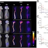
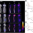
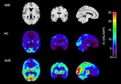 MRI (top panel) and coregistered F-18 AS2471907 PET images of a healthy control (middle panel) and an age- and sex-matched individual with AUD (bottom panel). Image courtesy of Neurobiology of Stress.
According to the findings, PET scans revealed 41.7% higher overall beta-11 HSD1 activity in the prefrontal-limbic circuit in the AUD group compared to the healthy control group, the researchers wrote.
Specifically, mean F-18 AS2471907 radiotracer values were higher in the AUD group by 47% in the amygdala, 36% in the caudate, 52% in the anterior cingulate cortex, 39% in the hippocampus, and 36% in the ventromedial prefrontal cortex (vmPFC).
“Our data suggest higher brain availability of the cortisol-regenerating enzyme beta-11 HSD1 in people with AUD, and that higher vmPFC beta-11 HSD1 availability is related to greater alcohol consumption,” the group wrote.
Ultimately, the researchers hypothesized that this higher beta-11 HSD1 availability in the brain may drive a blunted response to stress in individuals with AUD, which leads to increased vulnerability to excessive use. Yet they noted that the findings in this study are “first-in-kind” and that future studies will be needed to elucidate these mechanisms.
“These findings set the foundation for future hypotheses on mechanisms related to HPA axis function in this population,” the group concluded.
The full article is available here.


