PET imaging using a newly developed radiotracer has identified different patterns of brain tau pathology over time in early-onset versus late-onset Alzheimer’s disease patients, according to a study published February 1 in the Journal of Nuclear Medicine.
The study found that tau protein tangles showed rapid accumulation globally in early-onset patients and that this correlated with their verbal memory deterioration. Ultimately, the study validates F-18 PI-2620 PET as a biomarker that may be used to inform better treatment strategies for patients, noted lead author Minyoung Oh, MD, PhD, of the University of Ulsan in Seoul, Korea.
“These findings suggest that F-18 PI-2620 could be a potential biomarker for selecting tau-targeted therapies and monitoring their effects,” the group wrote.
The neuropathologic hallmarks of Alzheimer’s disease include extracellular accumulation of beta-amyloid plaques and accumulations of tau protein neurofibrillary tangles, the researchers explained.
However, the overall pathology of early-onset Alzheimer’s disease and late-onset Alzheimer’s disease patients is similar, making it difficult to distinguish between the two, they noted.
F-18 PI-2620 was first developed by researchers in Germany, and it has since been tested in studies that suggest it may be more accurate than other tau radiotracers used to diagnose Alzheimer’s disease patients based on its ability to bind more strongly to the target.
In this study, the group aimed to further test the approach to better understand the dynamic interactions between tau accumulation and beta-amyloid, neurodegeneration, and cognitive decline over time.
The team prospectively enrolled 52 participants (age, 69.7 ± 8.4 years; 18 men and 34 women): seven with normal cognition, 28 with mild cognitive impairment, and 17 with Alzheimer’s disease. All patients underwent F-18 PI-2620 PET imaging, as well as amyloid PET, MRI, and neuropsychologic tests at baseline and at follow-up after one year. In addition, the researchers classified the participants into early-onset (<65 years old) or late-onset (≥ 65 years old) groups.
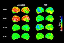
At baseline, mean global uptake of F-18 PI-2620 tracer (standard uptake values, or SUVs) was 1.04 in 15 patients with negative amyloid PET scans; 1.18 in 20 late-onset patients, and 1.54 in 17 early-onset patients.
 A graphical abstract illustrating the use of a new tau PET radiotracer in patients with Alzheimer's disease. Image courtesy of the Journal of Nuclear Medicine.
After one year, global SUVs increased by 0.05 (3.9%) in the late-onset patients and by 0.13 (8.4%) in the early-onset groups, while it remained unchanged in patients who were negative for beta-amyloid plaque.
Also, in the early-onset group, further analysis revealed these changes in SUV were strongly correlated with changes in patients’ verbal memory, according to the group.
“This study used F-18 PI-2620 PET to identify a tau deposition pattern and longitudinal accumulation, which differed between the late-onset and early-onset groups,” they wrote.
Ultimately, patients with Alzheimer’s disease are often classified into early-onset and late-onset groups based on age, with 65 being the cut-off age, the authors noted. This study provides a new understanding of differences among these groups based on tau brain pathology, which could open new avenues of research, they suggested.
“These findings suggest that [F-18]PI-2620 PET can be a biomarker to provide regional and chronologic information about tau pathology in the Alzheimer’s disease continuum,” the researchers concluded.
A link to the full article can be found here.