PET/CT is poised to become a key tool for imaging the coronary arteries – a clinical imaging exam that can reveal early warning signs of fatal conditions when patients present with symptoms of chest pain, experts say.
The “hybrid” scan – which combines molecular PET and anatomical CT images – may have its highest potential in visualizing early signs of so-called “perfusion deficits” (regions of obstructed blood flow), said Marcelo Di Carli, MD, a professor of radiology and medicine at Harvard Medical School, in a recent interview with AuntMinnie.com.
“[PET/CT] helps us differentiate a patient who has chest pain from obstructions of the coronary artery from another patient who may also have chest pain, but not coming from obstructed coronary arteries,” he said.
Di Carli discussed how PET/CT could revolutionize cardiac care.
Coronary artery disease (CAD) is the leading cause of mortality in the U.S. and accounts for approximately 610,000 deaths annually, with this number increasing, according to estimates. Obesity, physical inactivity, unhealthy eating, and smoking tobacco are risk factors.
In a recent editorial published in the Journal of Nuclear Cardiology, Di Carli, who is the editor in chief of the journal, described a study representing the largest validation to date of PET myocardial perfusion imaging (MPI) for detecting high-risk CAD.
In a cohort of 1,282 patients who underwent invasive coronary angiography for suspected disease, noninvasive PET MPI performed within six months identified large perfusion defects (>10% of the left ventricular myocardium) in 82% of the patients.
Di Carli noted that currently, SPECT MPI – also a nuclear medicine imaging exam, but one that requires higher radiation doses to patients and produces less resolution – remains the workhorse in the field. The game-changer could be the pending U.S. approval of new PET radiotracers, such as F-18 flurpiridaz, he said.
“The access to PET has been hindered primarily by the availability of radiopharmaceuticals for myocardial perfusion,” Di Carli said.
The approval of F-18 flurpiridaz could increase access to cardiac PET/CT exams.
Beyond PET MPI, there is increased use F-18 FDG PET/CT in the evaluation of inflammatory cardiomyopathies and large vessel vasculitis, with its role in evaluating patients with suspected or known cardiac sarcoidosis now integrated into national and internal guidelines, Di Carli noted.
Also recently, 11 medical societies jointly recommended PET/CT as the best method for diagnosing infections in people with ventricular assist devices, such as automatic defibrillators, with implants of these devices on the rise.
Finally, aside from developing the advanced machines used in PET/CT scans, industry is also stepping up to the plate. Earlier this month, three of the technology’s manufacturers formed a new coalition to promote federal policies that facilitate access to the imaging. Di Carli noted that manufacturers are aware that they need to bring down the cost of PET/CT scanners as use of the technology increases.
So, what does the near future look like for cardiac PET/CT? De Carli is quite optimistic.
“For patients with more advanced risk, older patients with a lot of cardiometabolic risk factors, I think PET will emerge as a very, very powerful technique,” he said.
Cardiac care centers are beginning to convert from older SPECT cameras to new PET/CT scanners.
You can listen to the full audio of the interview by clicking below.
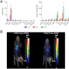


![A 53-year-old patient (patient number four) with a recurrent pituitary adenoma with extension of a cystic component of disease to the medial temporal lobe apparent on MRI (contoured in blue), and extension of disease to the left sphenoid bone and orbital apex apparent on [68Ga]Ga-DOTA-TATE (contoured in yellow).](https://img.auntminnie.com/mindful/smg/workspaces/default/uploads/2026/04/pituitary-tumor.QGsEnyB4bU.jpg?auto=format%2Ccompress&dpr=2&fit=crop&h=167&q=70&w=250)


