Proposed criteria for F-18 fluoromisonidazole (FMISO) PET imaging can help clinicians manage patients with head and neck cancer (HNC), according to a group at Memorial Sloan Kettering Cancer Center in New York City.
A team led by Rick Wray, MD, found that diagnostic criteria and a training protocol they developed improved interpretations of tumor hypoxia on F-18 FMISO-PET images among physicians with no experience.
The approach sets the stage for "broader application of F-18 FMISO imaging -- from academic to community practice -- and its use in future multicenter clinical trials," the group wrote. The study was published on September 12 in the Journal of Nuclear Medicine.
Tumor hypoxia is a prognostic and predictive cancer biomarker associated with resistance to chemotherapy, radiotherapy, and immunotherapy. In HNC, including HPV-positive (HPV+) oropharyngeal cancer, it is associated with a poor chemoradiotherapy outcome, the authors explained.
While F-18 FMISO-PET imaging has been used in research settings to image hypoxia for several decades, a major obstacle to its broader use is the lack of standardized interpretation criteria, the authors noted. To that end, the group developed and validated practical interpretation criteria.
The researchers randomly selected 123 images from patients with HPV+ oropharyngeal cancer enrolled in a previous phase II trial. Four independent nuclear medicine physicians with no experience in F-18 FMISO-PET read the scans. Interpretation by a fifth nuclear medicine physician with more than 20 years of experience was the reference standard.
The readers first received initial instruction via live web-based instruction and a group practice session to learn how to determine positive and negative scans. Next, they received dedicated training on more specific image characteristics, with their performances compared before and after the sessions.
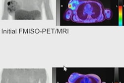
 A graphical abstract of the study. Image courtesy of the Journal of Nuclear Medicine.
After initial instruction, visual assessments by the readers produced a mean sensitivity and specificity of 77.3% and 80.9%, with fair inter-reader agreement (κ = 0.34), according to the findings. After dedicated training, mean sensitivity and specificity improved to 97.6% and 86.9%, with almost perfect agreement (κ = 0.86).
Additionally, the researchers determined whether defining a maximum standard uptake value (SUVmax) using tumor-to-background ratios could be helpful.
After initial instruction, quantitative assessment with an estimated SUVmax ratio threshold of more than 1.2 to define hypoxia positivity produced a mean sensitivity of 56.8% and a specificity of 95.9%, with substantial interreader agreement (κ = 0.66). After dedicated training, using an SUVmax of more than 1.2 improved sensitivity to 89.6%, whereas mean specificity remained high at 95.3%, with near-perfect interreader agreement (κ = 0.86), the group found.
"Nuclear medicine physicians without F-18 FMISO hypoxia PET reading experience demonstrate much improved interreader agreement with dedicated training using specific interpretation criteria," the researchers wrote.
Ultimately, based on several recent clinical trials, interest has grown in applying hypoxia F-18 FMISO-PET in managing patients with HNC treated with chemoradiation, the group wrote. The technique can help identify HPV+ oropharyngeal cancer patients who may benefit from de-escalation of therapy, for instance, as this type of cancer is hypoxia-negative, they wrote.
"There is growing interest in this application, and our work addresses the need for a standardized, reproducible method for F-18 FMISO PET/CT interpretation," the researchers concluded.
The full study is available here.


![A 53-year-old patient (patient number four) with a recurrent pituitary adenoma with extension of a cystic component of disease to the medial temporal lobe apparent on MRI (contoured in blue), and extension of disease to the left sphenoid bone and orbital apex apparent on [68Ga]Ga-DOTA-TATE (contoured in yellow).](https://img.auntminnie.com/mindful/smg/workspaces/default/uploads/2026/04/pituitary-tumor.QGsEnyB4bU.jpg?auto=format%2Ccompress&dpr=2&fit=crop&h=167&q=70&w=250)


