
F-18 FDG-PET/CT may be a useful diagnostic tool for detecting metastatic uveal melanoma, researchers have reported.
The finding is from a retrospective analysis of imaging results from 55 participants and suggests that F-18 FDG-PET/CT can play a crucial role in a group with low survival rates after metastases, noted corresponding author Nina Zhou, MD, of Peking University Cancer Hospital and Institute in Beijing, China, and colleagues.
“Early detection of metastatic uveal melanoma and intervention at the early stage of distant metastasis will be more conducive to improving patient survival,” the group wrote. The study was published October 24 in Cancer Imaging.
Uveal melanoma is a rare ocular tumor and is considered the most common primary malignant tumor in the adult eye. Although less than 5% of uveal melanoma patients are detected with metastasis at the initial diagnosis, up to 50% of patients will eventually develop distant metastasis over various lengths of time, the researchers explained.
At present, uveal melanoma has no established and effective treatments once metastasis occurs, with a median overall survival after initial diagnosis of metastases of about 12 months, they added.
F-18 FDG-PET is a widely used and effective scan for detecting metastatic cancer based on glucose metabolism in tumors, yet there is little evidence of its use in this patient group, the researchers noted.
To bridge the gap, the group analyzed results from 55 patients with uveal melanoma who underwent whole-body F-18 FDG-PET/CT scans (Biograph 64, Siemens Healthineers) during regular follow-up or for suspected recurrence after initial therapy between January 2011 and September 2024.
Among the 55 patients, 31 patients (56%) were confirmed to have metastasis through pathological or clinical follow-up. The average time from initial therapy to cancer recurrence was 30.7 months. Of these 31 patients, 13 had liver metastases only, 16 had both liver and extra-hepatic metastases, and two had extra-hepatic metastases only.
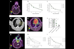
 Images of a 56-year-old female with recurrence of choriodal melanoma two years after left eye enucleation. (A) Maximum intensity projection (MIP) images showed multiple metastatic lesions. B-D Axial CT (B), PET (C), and fused PET/CT (D) images showed liver metastasis (green arrows) and splenic metastasis (yellow arrows). E-G images showed an L2 vertebral metastasis without density changes (red arrows). Cancer Imaging
In addition, among the 31 patients, a total of 270 lesions were confirmed by pathological or clinical follow-up, with F-18 FDG-PET/CT detecting 245 (90.7%) of these, the researchers reported.
The sensitivity of F-18 FDG-PET/CT in this setting was 90.3%, specificity was 100%, and accuracy was 94.5%, the group added.
“PET/CT is a very sensitive and specific tool for the early detection and localization of metastatic disease in patients with UM, particularly in bone and lymph node,” the researchers wrote.
Given the promising results, but noting the small sample size and inherent biases of retrospective analyses, validation of these results is warranted through prospective studies with larger cohorts, the group concluded.
The full study is available here.


![A 53-year-old patient (patient number four) with a recurrent pituitary adenoma with extension of a cystic component of disease to the medial temporal lobe apparent on MRI (contoured in blue), and extension of disease to the left sphenoid bone and orbital apex apparent on [68Ga]Ga-DOTA-TATE (contoured in yellow).](https://img.auntminnie.com/mindful/smg/workspaces/default/uploads/2026/04/pituitary-tumor.QGsEnyB4bU.jpg?auto=format%2Ccompress&dpr=2&fit=crop&h=167&q=70&w=250)


