PET/CT scans for staging patients with aggressive prostate cancer can also be used to screen for osteoporosis and vertebral fractures, researchers in France have reported.
The finding is from a feasibility study demonstrating that the low-dose CT component from F-18 fluorocholine (FCH) PET/CT scans is strongly correlated with standard stand-alone CT bone scans, noted lead author Astrid Dauchez, MD, of Cochin Hospital in Paris, and colleagues.
“This opportunistic screening approach could improve early osteoporosis detection in [prostate cancer] patients without requiring additional imaging, time or cost,” the group wrote. The study was published December 21 in Rheumatic and Musculoskeletal Diseases.
Androgen deprivation therapy (ADT), although effective in managing aggressive or advanced prostate cancer, can lead to bone loss and an increased fracture risk, the authors explained. While clinical guidelines thus recommend assessing osteoporosis risk prior to initiating ADT using gold standard dual-energy x-ray absorptiometry (DEXA), DEXA is infrequently performed in routine clinical practice, they added.
One promising alternative is measuring bone density using Hounsfield units (HU) on CT scans, the researchers wrote. Specifically, previous studies have shown that CT-derived lumbar trabecular attenuation (TA) strongly correlates with bone mineral density measured by DEXA, they noted.
In this study, the researchers hypothesized that the low-dose CT component of FCH-PET/CT scans could be used to opportunistically screen for osteoporosis and vertebral fractures.
The group retrospectively gathered data from 81 patients (mean age, 74.2 years old) with prostate cancer who underwent both FCH-PET/CT (primarily Discovery MI [n = 61], GE HealthCare) and stand-alone CT within six months at their hospital. Thirty-eight of the patients (46.9%) were current or former smokers, and 42 (51.9%) had received ADT prior to the FCH-PET/CT scan. Reasons for the PET/CT scans included initial staging (34.6%), biochemical recurrence (60.5%), and disease follow-up (4.9%).
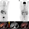
The investigators analyzed three vertebrae (L1, L2, and L3) per patient for a total of 243 vertebrae. TA values were comparable between the two imaging modalities, although attenuation values on stand-alone CT tended to be higher than those on FCH-PET/CT. Intraclass correlation coefficients (ICC) between TA values from FCH-PET/CT and stand-alone CT were 0.72, 0.7, and 0.67 for L1, L2, and L3, respectively (p < 0.001), reflecting good agreement, the researchers reported.
Further, the mean TA based on the low-dose FCH-PET/CT scans was 121 HU for L1, 122 HU for L2, and 118 HU for L3. Using a predefined threshold of 90 HU to define high-risk, 18 patients (22.2%) were considered at risk for vertebral fractures. Within this group, 44% had fractures, compared with 11.3% among patients with attenuation > 90 HU (p = 0.004). Finally, patients with fractures had significantly lower TA compared with those without fractures (mean, 87.8 vs. 127.5; p = 0.01).
“Our findings indicate that molecular imaging with FCH-PET/CT can provide reliable TA measurements for indirect [bone mineral density] estimation, comparable to conventional CT,” the group wrote.
While the association between TA and fracture risk requires validation in larger, prospective cohorts, the researchers hope this work will raise clinician awareness of bone health in prostate cancer management, particularly before initiation of ADT, and support preventive osteoporosis treatment strategies.
“This opportunistic strategy could raise clinician awareness of osteoporosis risk in [prostate cancer] and facilitate implementation of preventive treatments,” the group concluded.
The full study is available here.