
Prostate-specific membrane antigen (PSMA)-PET imaging shows promise as a tool for visualizing treatment responses in patients with hepatocellular carcinoma, researchers have reported.
In a head-to-head comparison in 88 patients treated with either locoregional therapy or immune checkpoint inhibitor therapy, PSMA-PET outperformed contrast-enhanced CT and MRI, noted lead author Ajith Antony, MD, of the Mayo Clinic in Rochester, MN, and colleagues.
“These findings provide the foundational support necessary to justify and design larger prospective trials to validate PSMA PET-based response against oncologic outcomes,” the group wrote. The study was published November 6 in the European Journal of Nuclear Medicine and Molecular Imaging.
PSMA is a protein highly expressed on prostate cancer cells, and PSMA-PET has become a standard imaging approach for evaluating patients with advanced prostate cancer. Yet PSMA is not limited to prostate cancer and can be found in other tumor entities, such as hepatocellular carcinoma (HCC), the authors explained.
In this study, the group’s primary objective was to determine whether PSMA-PET imaging can be interpreted consistently among radiologists and serve as a method for assessing HCC treatment responses.
Between July 2020 and December 2023, the researchers prospectively enrolled 13 patients with treatment-naive HCC who met the inclusion criteria for the study. Baseline gallium-68 (Ga-68) PSMA-11 PET/CT scans (Biograph Vision 600, Siemens Healthineers; Discovery MI, GE HealthCare) identified a total of 29 PSMA-avid target lesions (median lesion size: 2.1 cm).
Seven of the patients (53.8%; 10 lesions) underwent locoregional therapy (LRT) and six (46.2%; 19 lesions) received immune checkpoint inhibitor (ICI) therapy. Within approximately eight weeks, patients again had PSMA-PET scans, as well as contrast-enhanced CT and MRI scans.
Three radiologists independently assessed responses on the PSMA-PET images using a 4-point scale and on the CT and MRI scans using established, guideline-endorsed frameworks. The readers also independently assessed whether the lesions were “viable” or “nonviable” (binary status) on the PSMA-PET images to simulate clinical decisions on whether to modify or continue therapy.
According to the results, overall interreader agreement on whether patients had responded or not responded was almost perfect based on the PSMA-PET scans for both the 4-point qualitative scale (Gwet’s Agreement Coefficient, version 2 [Gwet’s AC2]: 0.86) and the binary viable/nonviable status [Gwet’s AC1]: 0.9).
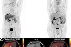
 Complete response to immune checkpoint inhibitor therapy on Ga-68 PSMA-PET/CT. Maximum intensity projection (MIP) images from an 83-year-old male with metastatic HCC treated with atezolizumab and bevacizumab. (A) Baseline pretreatment image demonstrates a large, intensely PSMA-avid lesion in the right hepatic lobe, along with multiple metastatic lesions throughout the abdomen and skeleton. (B) Follow-up scan performed four months after initiating therapy shows complete resolution of PSMA uptake in all hepatic and extrahepatic disease.European Journal of Nuclear Medicine and Molecular Imaging
“Our proof-of-concept study demonstrates that assessment of treatment response in HCC using a qualitative [Ga-68] PSMA PET/CT framework yields almost perfect inter- and intrareader reliability,” the researchers wrote.
The group noted that important next steps will be the development and validation of quantitative PSMA-PET response criteria in HCC. Such criteria could pave the way for PSMA-targeted theranostics in HCC, an area of active investigation, they added.
“It is envisioned that a baseline PSMA-PET would select the majority of HCC patients who are candidates for this monitoring and potential therapeutic pathway, while the subset of patients with non-avid disease would continue with standard-of-care imaging,” the researchers wrote.
The full study is available here.


![A 53-year-old patient (patient number four) with a recurrent pituitary adenoma with extension of a cystic component of disease to the medial temporal lobe apparent on MRI (contoured in blue), and extension of disease to the left sphenoid bone and orbital apex apparent on [68Ga]Ga-DOTA-TATE (contoured in yellow).](https://img.auntminnie.com/mindful/smg/workspaces/default/uploads/2026/04/pituitary-tumor.QGsEnyB4bU.jpg?auto=format%2Ccompress&dpr=2&fit=crop&h=167&q=70&w=250)


