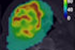
FDG-PET/CT and MRI show similar diagnostic accuracies in differentiating between malignant and benign soft-tissue tumors, but offer different attributes when imaging for bone tumors.
A study from Yeungnam University Hospital in Daegu, South Korea, found that FDG-PET/CT has a high negative predictive value with bone tumors, while MRI has a high positive predictive value in bone tumor imaging.
Through the retrospective study, researchers sought to determine FDG-PET/CT and MRI's efficacy with malignant tumors and benign lesions, given that certain tumors have no FDG uptake or distinguishing signal characteristics. The analysis reviewed 84 patients with primary musculoskeletal tumors. The patients had a mean age of 43 years. All patients underwent FDG-PET/CT and MRI before treatment.
"We used a cutoff barrier of 4.1 for maxSUV in soft-tissue tumors and 3.25 for maxSUV for bone tumors," added Dr. E. J. Kong during her presentation of the study at the annual SNM meeting. "All the results were confirmed through histologic diagnosis."
In comparing the results for differentiating between malignant and benign soft-tissue tumors, FDG-PET/CT and MRI showed similar efficacies in sensitivity, specificity, positive predictive value (PPV), and negative predictive value (NPV). (See chart below).
Differentiating between malignant and benign soft-tissue tumors
|
Kong noted that the maxSUV for malignant soft-tissue tumors was 13.2, while the maxSUV for benign soft-tissue tumors was 4.8.
In the study's analysis of differentiating between malignant and benign bone tumors, there were greater discrepancies, with FDG-PET/CT registering 49% specificity, compared with 94% for MRI. FDG-PET/CT scored higher than MRI in negative predictive value (100% versus 61%), while MRI showed prowess with positive predictive value (91% versus 68%).
Differentiating between malignant and benign bone tumors
|
Kong attributed FDG-PET/CT's low specificity with bone tumors to results from a segment of the patient sample that was less than 20 years old. In this under-20 group, she said, patients showed greater FDG uptake than patients older than 20 years.
"The younger patient group showed high sensitivity, but extremely low specificity," she said. "The older patient group showed relatively high sensitivity, positive predictive value, and negative predictive value."
Further comparison found a significant difference in maxSUV of benign (3.2 ± 3.0) and malignant (10.1 ± 7.5) bone tumors in patients older than 20 years, while specificity of FDG-PET/CT increased from 40% to 52% in the over-20-years sample.
Specificity was 39% in the younger group, compared with 52% in the older group. Positive predictive value was 68% in the under-20 segment, compared with 86% in the older group.
In conclusion, Kong noted that diagnostic accuracy in soft-tissue tumors did not significantly differ between FDG-PET/CT and MRI, but FDG-PET/CT performed with a greater negative predictive value.
MRI offered a greater positive predictive value in bone tumors than FDG-PET/CT, as well as significantly increased specificity for bone tumors in patents older than 20 years.
By Wayne Forrest
AuntMinnie.com staff writer
September 3, 2008
Related Reading
FDG-PET/CT trumps bone scintigraphy for young Hodgkin's lymphoma patients, June 30, 2008
FDG-PET changes treatment plans for glioma patients, June 26, 2008
Open-source software delivers 3-way (PET/CT/MR) image fusion, April 25, 2008
SPECT/CT offers on-target diagnosis in noncancerous bone disease, March 23, 2007
Copyright © 2008 AuntMinnie.com


