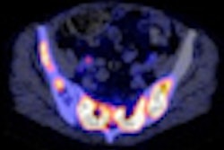
The use of ultralow-dose CT scans for attenuation correction and respiratory gating of PET scans is feasible, concluded a recent study published in Physics in Medicine and Biology. Doses an order of magnitude lower than existing low-dose CT protocols on PET/CT scanners were achieved.
"The methods we have developed will directly benefit patients receiving extended-duration CT scans as part of a PET/CT acquisition protocol, most likely those with lung and abdominal cancers, where respiratory motion is the greatest," explained medical physicist and senior author Paul Kinahan, from the University of Washington.
The value of quantitative analysis of F-18 FDG PET images in oncology, using parameters such as standardized uptake value (SUV), is increasingly of interest. Such parameters are potential indicators of prognosis early on in a patient's treatment course. However, variation in photon attenuation with tissue depth and the blurring effects of breathing motion introduce inaccuracies that must be corrected for if such information is to be exploited fully (Phys Med Biol, January 21, 2012, Vol. 57:2, pp. 309-328).
CT-based attenuation correction and respiratory gating are popular solutions to resolve these problems. Respiratory-gated CT is phase-matched to correct for attenuation in gated PET data, which in turn is used to separate photons from different respiratory phases. However, this process involves extended, continuous CT data acquisition over several breathing cycles, which can result in effective doses to the patient as high as 16 mSv.
Reducing the dose
The researchers set out to identify whether ultralow-dose CT scans could be used to perform attenuation correction and respiratory gating in PET data. Ultralow-dose CT would enable the extended CT acquisitions necessary for gating, while minimizing dose to the patient. They exploited the fact that such scans, while exhibiting lower signal-to-noise ratios and inferior image quality compared to diagnostic scans, are nevertheless adequate for the purposes of attenuation correction and respiratory gating.
"A key enabler was the recognition that if a diagnostic-quality CT image is not needed, we can significantly reduce the CT radiation dose without degrading the accuracy of the PET image," explained Kinahan. "This led to a fun part of this project: discussions with CT experts in industry and academia to convince them that we can relax the requirements for CT image quality in some conditions. At first they were skeptical, quite reasonably, but once convinced they became very enthusiastic about the concept."
The researchers explored several dose reduction strategies using simulations and phantom measurements. Simulation software included a Monte Carlo package, CATDOSE, which enabled CT dose to be estimated. Strategies included optimization of scanning parameters such as tube voltage and filtration. The researchers also used indirect approaches to compensate for the lower signal-to-noise ratios associated with ultralow-dose CT, reducing image noise with sinogram smoothing and clipping.
Simple alteration of CT acquisition parameters was enough to achieve ultralow-dose CT, while at the same time producing attenuation corrections of acceptable accuracy in accompanying PET scans. For example, scanning with a tube voltage of 140 kVp and 1 mm of copper filtration to increase photon penetration resulted in a CT absorbed dose of 0.14 mGy at the central slice of the scan. This was a threefold reduction compared to scanning with 80 kVp and no filtration.
Furthermore, sinogram smoothing was found to reduce CT noise in ultralow-dose scans, enhancing the capacity of the CT data to correct for attenuation in accompanying PET data. Indirectly, this strategy was demonstrated to reduce CT dose by up to a factor of three. The combined effects of these methods indicated the effective dose from the CT component of the PET/CT scan could be reduced from 16 mSv to less than 3 mSv without degrading PET image quality.
The findings have potential dose-saving benefits for patient populations other than oncology. "The ultralow-dose CT acquisitions can be used in any protocol where a high-dose diagnostic CT is not needed as part of a PET/CT scan, including pediatric imaging," Kinahan told medicalphysicsweb.
© IOP Publishing Limited. Republished with permission from medicalphysicsweb, a community Web site covering fundamental research and emerging technologies in medical imaging and radiation therapy.


