NEW ORLEANS - Cardiac MRI can be used as a tool to assess the impact of statin therapy in reducing atherosclerotic plaque, possibly earlier than other modalities, according to a paper presented at the American College of Cardiology meeting this week.
Statin drugs have emerged as powerful weapons in treating atherosclerosis, the artery-clogging condition in which fatty plaques accumulate. Studies indicate that statin therapy can reduce low-density lipoprotein (LDL) cholesterol levels by 30%, with a corresponding 30% reduction in subsequent cardiac events, according to Dr. Joao Lima of Johns Hopkins University in Baltimore.
The theory is that the beneficial impact of statins is due to plaque regression, in which plaques grow smaller after statin therapy. Imaging techniques such as ultrasound and coronary angiography have been used to track plaque regression, but most of these studies have measured changes a year after therapy began.
At the same time, pathological studies have indicated that statin therapy begins to show benefits as soon as three months after therapy starts. Could cardiac MRI's superior spatial resolution enable clinicians to measure plaque regression earlier than has been demonstrated by other imaging techniques? Lima's group decided to put the modality to the test.
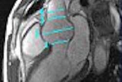
For its study, the group used a transesophageal probe with three surface coils to image atherosclerosis in the aorta. They used a composite of six slices to generate a volume, and used plaque area and plaque volume as the indices by which plaque regression would be measured. The study involved 29 patients with advanced atherosclerosis (85% had peripheral vascular disease) who were given simvastatin (Zocor, Merck, Whitehouse Station, NJ). The patients were followed at six-month intervals.
As expected, the statins had the desired effect on cholesterol levels: Total average cholesterol fell from 116 LDL to 84 LDL, Lima said. And at 12 months, MRI measured plaque regression of 30%. But the modality also demonstrated plaque regression at six months, Lima said, and the reduction in plaque volume directly correlated with the reduction in LDL cholesterol.
Those patients with LDL scores that had dropped by 30 points showed significant reduction in plaque volume. Patients with little reduction in LDL had much less reduction in plaque volume, and some patients who didn't change had a progression of stenosis.
The Hopkins group's results were based on imaging of the greater vessels of the heart, and Lima said the next step is to see if they can show the same results in the coronary arteries. Besides MRI, multislice CT could also prove of value. Lima's group is working with a new 32-slice model that John Hopkins has acquired.
Lima believes that the research indicates that MRI and perhaps CT could add to their growing role in cardiac imaging by becoming useful tools for measuring statin therapy.
"Statin-induced plaque regression and reverse remodeling may occur as early as six months after onset of statin therapy and is in proportion to lipid lowering," he said. "MRI and CT have the potential to provide simultaneous assessment of plaque burden and luminal patency in different arterial territories, including the coronaries."
By Brian CaseyAuntMinnie.com staff writer
March 7, 2004
Related Reading
New MRI approach may differentiate beta-blocker candidates with Marfan syndrome, March 2, 2004
Myocardial perfusion measured by MRI useful in detecting heart disease, August 4, 2003
Transesophageal MRI improves evaluation of aortic plaque, April 4, 2003
Copyright © 2004 AuntMinnie.com

