U.K. investigators found that MRI can have a positive impact on the management of patients with ankle disorders. Dr. Philip Bearcroft and colleagues performed a prospective observational study in 91 consecutive patients. The group, from Addenbrooke's Hospital and the Institute of Public Health, both in Cambridge, also assessed MRI's impact on the orthopedic specialist's diagnosis as well as diagnostic certainty.
"A visual analogue scale (VAS) was used to record clinician's estimates of preimaging probability for the most common diagnosis," the authors wrote. These included osteoarthritis or disorders of the Achilles. "Once the MRI examination had been performed, the findings and images were presented to the surgeon at a meeting with the radiologist" (American Journal of Roentgenology, November 2006, Vol. 187:5, pp. 1327-1331).
The surgeon completed two forms -- before and after imaging results -- that recorded the diagnosis, diagnostic certainty on VAS, and proposed management. The MRI protocol on a 1.5-tesla (LX Horizon EchoSpeed, GE Healthcare, Chalfont St. Giles, U.K.) or 1-tesla (Gyroscan, Philips Medical Systems, Andover, MA) system included a coronal T1 sequence and gradient-echo T2* sequences.
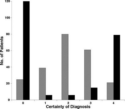
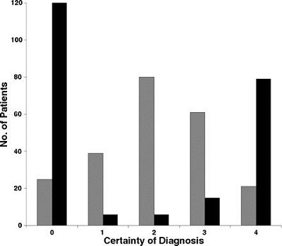
The study results showed a significant reduction in the possible diagnoses after MR imaging with an increase in diagnostic certainty on the part of the surgeon in 71% of the patients. Nineteen new diagnoses were made after MRI and 84% of these were considered definite or probable.
 |
| Bar graph shows changes in diagnostic certainty after MRI for all diagnoses under consideration. Gray bars = before MRI, black bars = after MRI. An entry in the zero column represents postoperative diagnosis of normal. Bearcroft PWP, Guy S, Bradley M, and Robinson F, "MRI of the Ankle: Effect on Diagnostic Confidence and Patient Management" (AJR2006; 187:1327-1331). |
Management plans changed in 35% of all patients with 14% of the 65 patients scheduled for surgery undergoing nonsurgical therapy. In six cases in which open surgery remained the treatment of choice, the surgeon stated that MRI results did inspire him to alter his surgical approach.
"The fact that the surgeon thought that diagnostic confidence either has been substantially improved by or had depended on the results of the MRI in 66% of the patients is a testament to the usefulness of ankle MRI in this practice," the authors stated.
While surgery couldn't be avoided in a substantial number of patients (54 of 65), other more sophisticated MR techniques, such as MR arthrography, may have impacted the management strategy to a greater degree, they added. The strength of conventional MRI in this setting was that it could effectively cut down on the number of possible ankle disorder diagnoses.
By Shalmali Pal
AuntMinnie.com staff writer
March 12, 2007
Related Reading
High signal on T2 MR warns of poor healing after Achilles tendon repair, December 22, 2006
US overcomes x-ray's limits in pediatric ankle fractures, January 23, 2006
Copyright © 2007 AuntMinnie.com


