(Booth 5529) GE Healthcare of Chalfont St. Giles, U.K., will bring the newest advances for its Signa MRI product line, from technology hardware to elite clinical applications, to RSNA 2008.
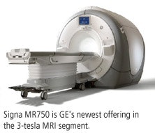
GE will display its Signa MR750 3-tesla system, which features GE's advanced thermal management system and is designed for as much as 60% more anatomical coverage and resolution unit per time.

GE also will highlight advanced clinical applications made available on the Signa MR750 platform. Lava-Ideal is a dual-echo acquisition technique for detailed, 3D abdominal images in one breath-hold. Clinicians can select output image types, such as in-phase, opposed-phase, water, and fat, to produce four image contrasts with only one scan. Clinicians can perform a complete liver exam in 15 minutes, according to GE.
Vibrant-Ideal is another new application designed for fat-free breast imaging with enhanced spatio-temporal resolution. Vibrant-Ideal catches the shortest in- and out-of-phase echoes to keep scan times comparable to single-echo acquisitions, even though twice the amount of data is collected.
Vibrant-Ideal also is designed to optimize acquisition with a high signal-to-noise ratio for acquiring high-quality water and fat images, allowing users to prescribe thinner slices for imaging with high spatial resolution.
GE also will unveil Propeller 2.0 for all neuro imaging planes with the implementation of the company's "no phase wrap" (NPW) technique. According to the company, NPW allows scans that are virtually free of ghost and motion artifacts in sagittal, coronal, axial, and oblique planes.
GE's exclusive 3D SWAN technique is designed to capture and delineate small vessels and microbleeds, as well as large vascular structures and deposits of iron and calcium in the brain. 3D SWAN's multiecho acquisition technique and postprocessing algorithm provide wider tissue bandwidth, two to four times more signal-to-noise ratio, and more contrast within clinically relevant scan times.
3D SWAN also may aid clinicians in diagnosing, among other things, patients with ischemic and cerebral brain diseases, arteriovenous malformations, and neurodegenerative diseases.


