Cardiovascular MRI has revealed that stress cardiomyopathy may have broader clinical characteristics than previously reported, including younger patients, men, and patients without an identifiable stressful cause, according to a study in the July 20 issue of the Journal of the American Medical Association.
In addition, when cardiovascular MRI is used when a patient first presents with possible symptoms, the exam may provide relevant functional and tissue information to confirm or rule out a diagnosis of stress cardiomyopathy, according to a research team from the University of Leipzig in Germany.
Stress cardiomyopathy is a form of acute heart failure that is triggered by stressful events and primarily affects postmenopausal women. It is characterized by acute and profound but reversible left ventricular (LV) dysfunction, according to the researchers.
"Various aspects of [stress cardiomyopathy's] clinical profile have been described in small single-center populations, but larger, multicenter datasets have been lacking so far," wrote lead study author Dr. Ingo Eitel and colleagues. "Furthermore, it remains difficult to quickly establish diagnosis on admission."
The study was conducted at seven tertiary care centers in Europe and North America between January 2005 and October 2010. A total of 256 patients with stress cardiomyopathy were assessed at the time of presentation at the centers and again one to six months after the acute event (JAMA, July 20, 2011, Vol. 306:3, pp. 277-286).
Patients had an average age of 69 years, and 227 (89%) were women. Among the female patients, 207 (81%) were postmenopausal, while 20 (8%) were 50 years of age or younger. In 182 patients (71%), the researchers noted a significant stressful event less than 48 hours before presentation could be identified. The events included emotional stress in 30% of patients and physical stress in 41% of the patients.
Electrocardiograms (ECGs) revealed abnormalities in 226 patients (87%), while coronary angiography showed healthy coronary arteries in 193 patients (75%).
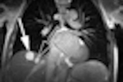
Cardiovascular MRI detected ballooning patterns in the heart muscle with moderate to severe reduction of LV function in all patients, with four distinct patterns of regional ventricular ballooning.
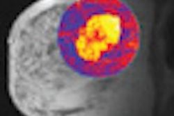
"Stress cardiomyopathy was accurately identified by cardiovascular MR using specific criteria: a typical pattern of LV dysfunction, myocardial edema, absence of significant necrosis/fibrosis, and markers for myocardial inflammation," the authors wrote. "Follow-up cardiovascular MR imaging showed complete normalization of LV ejection fraction and inflammatory markers in the absence of significant fibrosis in all patients."
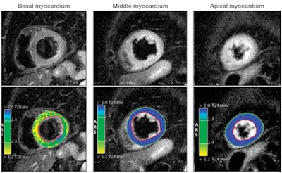 |
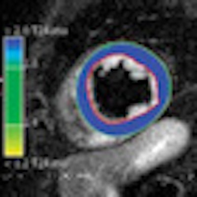
| T2-weighted images demonstrate normal signal intensity (SI) of the basal myocardium but global edema of the mid and apical myocardium. Computer-aided SI analysis (bottom row) of the T2-weighted images with color-coded display of relative SI normalized to skeletal muscle confirms the presence of global mid and apical edema (blue indicates an SI ratio of myocardium to skeletal muscle of 1.9 or greater, indicating edema; green/yellow indicates a normal SI ratio of < 1.9). Outlines of regions of interest are manually drawn around the myocardium (red contour = subendocardial border; green contour = subepicardial border) and within the skeletal muscle (contour not shown). Copyright © 2011 American Medical Association. All rights reserved. |
Eitel and colleagues also found that two-thirds of the patients had a clearly identifiable preceding stress trigger, compared with previous research where the preceding emotional or physical factors were as great as 89%.
"Thus, our large multicenter cohort demonstrates that the absence of an identifiable stressful event does not rule out the diagnosis, and, hence, precipitating mechanisms may be more complex, such as involvement of vascular, endocrine, and central nervous systems," they noted. "Such clinical heterogeneity could contribute to ambiguity in the recognition of stress cardiomyopathy and thereby affect potential management strategies."
Greater awareness and recognition of a broad clinical profile of stress cardiomyopathy -- as demonstrated in this study -- is "mandatory for correct diagnosis and treatment among patients with suspected stress cardiomyopathy," according to the authors.


