Researchers at Washington University School of Medicine in St. Louis have developed an MRI technique that provides access to brain landmarks previously available only at autopsy, according to a study published August 10 in the Journal of Neuroscience.
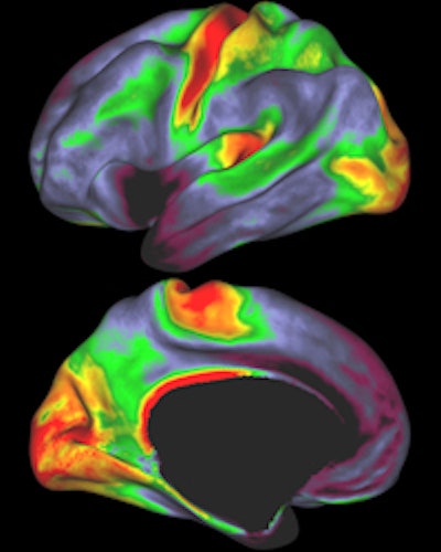
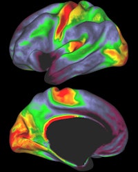 Red and yellow indicate regions with high myelin levels, while blue, purple, and black areas have low myelin levels. Image courtesy of David Van Essen, PhD.
Study co-author David Van Essen, PhD, head of the department of anatomy and neurobiology at Washington University, said the myelin maps will provide important insight into where certain parts of the brain end and others begin.
Access to detailed myelination maps in humans and animals will also help efforts to understand how the brain evolved and how it functions.
The technique was developed through the Human Connectome Project, a $30 million, five-year effort to map the brain's connectivity. The project is led by Washington University and the University of Minnesota.


