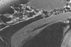
Researchers using a new MRI technique have found that the presence of particular fatty acids in the breast may be a useful indicator of cancer in postmenopausal women, according to a study published online June 7 in Radiology.
The role of body fat composition in breast cancer development has been thoroughly studied, using measures such as body mass index (BMI) and dietary fat intake. But the exact mechanism behind the increased risk of breast cancer in postmenopausal women with higher BMIs is not clear, wrote the group led by Melanie Freed, PhD, of NYU Langone Medical Center (Radiology, June 7, 2016).
For this preliminary investigation, Freed's team developed an MR method called gradient-echo spectroscopic imaging that estimates fractions of different types of fat in breast adipose tissue, such as monounsaturated fatty acids, polyunsaturated fatty acids, and saturated fatty acids. The technique is part of a clinical breast MRI exam and can be easily incorporated into standard workflow.
"Studies that evaluated the effect of the composition of dietary fat intake and cancer risk showed conflicting results," the group wrote. "In addition, adipose tissue composition differs from dietary intake values because some fatty acids can be manufactured in the body. Therefore, direct measures of fat composition may correlate more closely with cancer risk."
And imaging is a key resource in this regard, corresponding author Sungheon Kim, PhD, told AuntMinnie.com.
"Most of the studies out there that have tried to explore the link between fat and breast cancer haven't used imaging to do so," he said. "We wanted to use a noninvasive, in vivo method to address the issue."
What kind of fat?
The study included 89 women, 58 of whom were premenopausal and 31 of whom were postmenopausal. The women underwent MRI between July 2013 and September 2014. Each subject's height and weight were measured at the time of the exam, and their body mass index was calculated. In addition, the researchers determined each woman's breast cancer status: 49 had benign breast tissue, 12 had ductal carcinoma in situ, and 28 had invasive ductal carcinoma.
Freed and colleagues found that postmenopausal women with invasive ductal carcinoma had a greater proportion of saturated fatty acids and a lower proportion of monounsaturated fatty acids in their breast tissue compared to postmenopausal women with benign breast tissue. Also, among women with benign lesions, postmenopausal women had higher polyunsaturated fatty acids and lower saturated fatty acids in their breasts than premenopausal women.
Surprisingly, the researchers did not find a correlation between BMI and the fat composition of breast tissue, even though a higher BMI is considered a risk factor for developing the disease, according to the group.
"BMI and fat composition may affect cancer risk via independent mechanisms," they wrote.
More research needed
The study cohort included women scheduled for high-risk screening or follow-up or suspected of having cancer, Freed and colleagues noted. Because of this, more research is needed in low-risk patients to see if similar differences are found. Also, the study data were unclear on whether there was a causal relationship between saturated fatty acids and monounsaturated fatty acids and the development of invasive breast cancer.
"If there is a causal relationship, it may point to internal biologic factors as more important to invasive cancer development than dietary intake," they wrote.
The researchers plan to further investigate the relationship between higher levels of saturated fat and tissue estrogen levels and cancer development, Kim said.
"Ours was a preliminary study, to demonstrate that the technique works," he said. "But it would be great if these findings could lead to identifying more breast cancer risk factors."


