Based on alterations in cerebral blood flow (CBF) in key brain regions, MR images may provide an early warning sign of abnormal brain development in very premature infants, according to a prospective study published online December 3 in the Journal of Pediatrics.
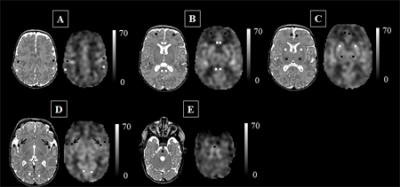
The researchers used arterial spin labeling (ASL) MRI to label water and map how blood flows through infants' brains to detect which regions do or do not receive adequate blood supply. With ASL-MRI, scans can be performed without a contrast agent because water from arterial blood illuminates the path traveled by cerebral blood. The technique also appears to make an abnormality more visible than conventional imaging does.
 Catherine Limperopoulos, PhD.
"During the third trimester of pregnancy, the fetal brain undergoes an unprecedented growth spurt," senior author Catherine Limperopoulos, PhD, director of the Developing Brain Research Laboratory in Washington, DC, said in a press release. "To power that growth, cerebral blood flow increases and delivers the extra oxygen and nutrients needed to nurture normal brain development. In full-term pregnancies, these critical brain structures mature inside the protective womb where the fetus can hear the mother and her heartbeat, which stimulates additional brain maturation. For infants born preterm, however, this essential maturation process happens in settings often stripped of such stimuli."
The challenge, she added, is how to capture what goes right or wrong in the developing brains of these fragile newborns.
The researchers studied 98 preterm infants who were born between June 2012 and December 2015, were younger than 32 gestational weeks at birth, and weighed less than 1,500 g. Those subjects were matched by gestational age with 104 infants who successfully completed their term (J Pediatr, December 3, 2017).
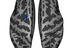
Compared with the full-term infants, the preterm infants had a significantly decreased volume of blood in several regions:
- The insula, which is critical to experiencing emotion.
- The anterior cingulate cortex, which is associated with cognitive processes.
- The auditory cortex, which helps process sound.
 Images show cerebral blood flow maps for a preterm infant scanned at term age without evidence of brain injury. The insula (black arrows in panel D) may be particularly vulnerable to stresses outside the womb. Images courtesy of Bouyssi-Kobar et al., Journal of Pediatrics.
"The ongoing maturation of the newborn's brain can be seen in the distribution pattern of cerebral blood flow, with the greatest volume of blood traveling to the brainstem and deep gray matter," said lead author Marine Bouyssi-Kobar. "Because of the sharp resolution provided by ASL-MRI, our study finds that in addition to the brainstem and deep gray matter, the insula and the areas of the brain responsible for sensory and motor functions are also among the most oxygenated regions."
The findings underscore the importance of these brain regions in early brain development, Bouyssi-Kobar added. In preterm infants, the insula may be especially vulnerable to conditions outside the womb.


