
Approximately half of patients considered to have a low risk of developing atherosclerosis showed at least one narrowed artery on whole-body MR angiography (MRA) exams in a study published online May 1 in Radiology.
The results confirm MRA's feasibility as an imaging method for detecting early atherosclerotic disease in individuals believed to have a low to intermediate risk of cardiovascular events, according to study co-author Dr. Graeme Houston from the University of Dundee in Dundee, Scotland.
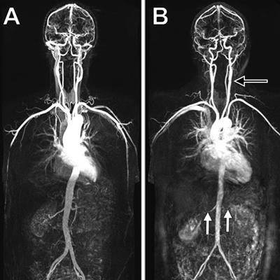
 Whole-body MRA shows no evidence of disease in a 45-year-old woman (A). MRA in a 53-year-old woman (B) discovered extensive disease, including left carotid bulb stenosis (black arrow), bilateral renal artery stenosis (white arrows), and an occluded right superficial femoral artery (arrowhead). Image courtesy of RSNA and Radiology.
"This approach could stratify individuals for the presence of disease burden, which could inform further preventative therapy in the future," he said in a statement.
Atherosclerotic disease often leads to cardiovascular disease -- the leading cause of death in the U.S. Early intervention can slow or reverse disease progression, but standard imaging techniques for early detection focus on specific artery segments, potentially missing evidence of disease elsewhere in the body, according to the authors.
Houston and colleagues used whole-body MRA to assess 31 arterial segments in 1,513 people who were asymptomatic for atherosclerosis. The subjects had an average age of 53.5 years and were considered to have less than 20% risk of developing cardiovascular disease within 10 years (Radiology, May 1, 2018).
Using whole-body MRA, the researchers detected early atherosclerotic disease throughout the subjects' bodies and interpreted 99.4% of the potentially analyzable arterial segments. They found that almost half of the participants had at least one narrowed vessel and more than 25% had more than one narrowed vessel.
"This is surprising, given that the study group was made up of asymptomatic individuals without diabetes who had low to intermediate risk of future cardiovascular events by standard risk factor assessment," Houston said.
The researchers said they plan to continue with follow-up studies to determine if there are any links between whole-body MRA findings and long-term health outcomes.
"The key advantages of MRA include the whole-body approach, which detects systemic disease that would be missed by modalities assessing single vascular sites," Houston said. "The results offer a validated quantitative score of atherosclerotic burden, and the technique does not use ionizing radiation, which is an advantage over CT angiography."


![Overview of the study design. (A) The fully automated deep learning framework was developed to estimate body composition (BC) (defined as subcutaneous adipose tissue [SAT] in liters; visceral adipose tissue [VAT] in liters; skeletal muscle [SM] in liters; SM fat fraction [SMFF] as a percentage; and intramuscular adipose tissue [IMAT] in deciliters) from MRI. The fully automated framework comprised one model (model 1) to quantify different BC measures (SAT, VAT, SM, SMFF, and IMAT) as three-dimensional (3D) measures from whole-body MRI scans. The second model (model 2) was trained to identify standardized anatomic landmarks along the craniocaudal body axis (z coordinate field), which allowed for subdividing the whole-body measures into different subregions typically examined on clinical routine MRI scans (chest, abdomen, and pelvis). (B) BC was quantified from whole-body MRI in over 66,000 individuals from two large population-based cohort studies, the UK Biobank (UKB) (36,317 individuals) and the German National Cohort (NAKO) (30,291 individuals). Bar graphs show age distribution by sex and cohort. BMI = body mass index. (C) After the performance assessment of the fully automated framework, the change in BC measures, distributions, and profiles across age decades were investigated. Age-, sex-, and height-adjusted body composition reference curves were calculated and made publicly available in a web-based z-score calculator (https://circ-ml.github.io).](https://img.auntminnie.com/mindful/smg/workspaces/default/uploads/2026/05/body-comp.XgAjTfPj1W.jpg?auto=format%2Ccompress&fit=crop&h=100&q=70&w=100)


![Overview of the study design. (A) The fully automated deep learning framework was developed to estimate body composition (BC) (defined as subcutaneous adipose tissue [SAT] in liters; visceral adipose tissue [VAT] in liters; skeletal muscle [SM] in liters; SM fat fraction [SMFF] as a percentage; and intramuscular adipose tissue [IMAT] in deciliters) from MRI. The fully automated framework comprised one model (model 1) to quantify different BC measures (SAT, VAT, SM, SMFF, and IMAT) as three-dimensional (3D) measures from whole-body MRI scans. The second model (model 2) was trained to identify standardized anatomic landmarks along the craniocaudal body axis (z coordinate field), which allowed for subdividing the whole-body measures into different subregions typically examined on clinical routine MRI scans (chest, abdomen, and pelvis). (B) BC was quantified from whole-body MRI in over 66,000 individuals from two large population-based cohort studies, the UK Biobank (UKB) (36,317 individuals) and the German National Cohort (NAKO) (30,291 individuals). Bar graphs show age distribution by sex and cohort. BMI = body mass index. (C) After the performance assessment of the fully automated framework, the change in BC measures, distributions, and profiles across age decades were investigated. Age-, sex-, and height-adjusted body composition reference curves were calculated and made publicly available in a web-based z-score calculator (https://circ-ml.github.io).](https://img.auntminnie.com/mindful/smg/workspaces/default/uploads/2026/05/body-comp.XgAjTfPj1W.jpg?auto=format%2Ccompress&dpr=2&fit=crop&h=167&q=70&w=250)


