MRI has identified parts of the brain needed to remember words -- and how those parts may be affected by epilepsy, according to a study published March 19 in Brain Communications.
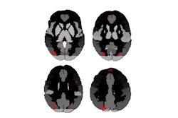
A team led by Giorgio Fiore, MD, of University College London (UCL), found that shrinkage in the prefrontal, temporal, and cingulate cortices, and the hippocampus areas of the brain was linked to difficulty remembering words -- indicating how the network involved in creating and storing word memories is dispersed and offering insight into how to best plan surgery for epilepsy treatment.
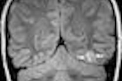
"Detailed MRI brain scans are used to find out the causes of epilepsy and can show if any parts of the brain are shrunken," said corresponding author John Duncan, MD, also of UCL, in a university statement. "By measuring the parts of the brain that are shrunken and how well a person can remember words, we can work out which parts … are used for making and storing memories. In addition, if medication has not stopped seizures from occurring, this helps us to guide neurosurgical treatment for a person's epilepsy, to avoid damaging the parts of the brain that are needed for memory to work well."
The team noted that "europhysiological and lesion studies have shown that increased activity of the prefrontal cortices is associated with better verbal memory encoding, and patients with lesions in these regions have verbal learning impairment." But cortical regions studied have been limited.
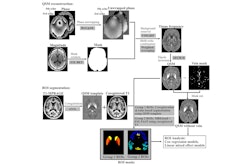
To address the knowledge gap, Fiore and colleagues conducted a study that included 84 people with temporal lobe epilepsy and hippocampal sclerosis and 43 people without these conditions. Patients underwent high-resolution MRI scans to measure the size and shape of different parts of the brain, including the cerebral cortex (which is responsible for thinking, memory, attention, perception, awareness, and language) and specific areas within the hippocampus (which helps with learning, memory, and spatial navigation). Study participants then underwent tests taken from the Adult Memory and Information Processing Battery to assess their verbal memory and the investigators compared the test scores to the sizes of different brain areas as visualized on MRI.
Overall, the team found that smaller sizes in brain areas such as the prefrontal, temporal, and cingulate cortices, and parts of the hippocampus were linked to worse memory for words in people with temporal lobe epilepsy.
The results could help better guide surgery for epilepsy, Fiore said in the UCL statement.
"[Our] research is important as it helps us to understand how memory may fail and may help guide the designing of neurosurgical operations for epilepsy that will not make memory worse," he noted.
The full study can be found here.