

Functional MRI (fMRI) is further revealing changes in the brain caused by repetitive blast exposure and highlights the need to rethink "mild" brain injuries across society, according to research published April 1 in Radiology.
A team led by neuroradiologist Andrea Diociasi, MD, from Mass General Brigham in Boston studied 220 U.S. military special operations forces members and found that when standard MRI exams appeared normal, resting-state fMRI maps obtained from functional connectivity (FC) multivariate patterns analysis, along with volumetric analysis, showed distinct differences between brain regions of interest, depending on blast exposure.
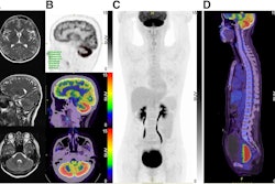
 Functional MRI (fMRI) scans depict the merged regions of interest that showed significant differences between the two datasets of high and low blast exposure groups. The clusters involved the bilateral superior and inferior lateral occipital cortex, the frontal medial cortex, left superior frontal gyrus, and precuneus. Image courtesy of the RSNA.
For the prospective study, participants in the Comprehensive Brain Health and Trauma (ComBHaT) program underwent psychodiagnostics and neuroimaging evaluation using structural and resting-state fMRI. Researchers divided participants into two datasets (n = 161) and an independent validation set (n = 51), which they further divided into low and high blast-injury exposure groups.
Only those with MRI reports classified as normal by a board-certified neuroradiologist were included, ensuring the absence of structural abnormalities, the authors noted. Eight participants were excluded due to motion artifacts on fMRI images or incomplete imaging protocols.
From the final cohort of 212, those with higher blast exposure showed differences in functional connectivity in the superior and inferior lateral occipital cortex (LOC), frontal medial cortex, left superior frontal gyrus, and precuneus (p = 0.001 to 0.04), compared with the low-exposure group, the authors noted.
"With our larger cohort, we confirmed altered connectivity ... likely due to recurrent minor blast injuries," the team noted. Following the fMRI analysis, they conducted a volumetric study for each group.
Participants with higher exposure to significant explosives showed increased volume in the left and right superolateral occipital cortices. In particular, the higher large explosive exposure group in dataset 1 (n = 161) showed significantly greater volumes in the right superior LOC compared to the lower-exposure group (19,431 mm3 ± 210.0 vs. 19,166 mm3 ± 190.4, respectively; p = 0.016) and in the left superior LOC compared with the lower exposure group (19,550 mm3 ± 209.0 vs. 18,911 mm3 ± 187.7; p = 0.018).
The high-exposure group also scored higher on the Neurobehavioral Symptom Inventory and Posttraumatic Stress Disorder Checklist for the Diagnostic and Statistical Manual of Mental Disorders. Clinical scores from both were inversely correlated with FC in regions linked to cognition, attention, and emotional regulation, including the LOC, superior parietal lobule, precuneus, and default mode networks (r = −0.163 to −0.384; p < 0.001 to 0.04), the team reported.
While they acknowledged the study's limitations, Diociasi and colleagues said that their method may bridge the divide between clinical observations and imaging outcomes in participants exposed to blasts. Future studies should use advanced models integrating multimodal imaging to enhance the detection of subtle blast-induced neural changes, the authors added.
“Repeated trauma seems to weaken the brain’s internal communication," stated Diociasi for the RSNA, noting that across very different populations, the same pattern emerges: mild but repetitive trauma can have lasting effects. “The broader implication is that we need to rethink how we view ‘mild’ brain injuries, not just in soldiers, but across society."
"The results were not surprising, as it is increasingly clear that even mild, repetitive trauma can affect both brain function and morphovolumetric metrics," Diociasi told AuntMinnie in an email. "Our study builds upon numerous previous findings, particularly in military populations and others exposed to repeated trauma.
"Our next goal is to expand the research using additional modalities to gain deeper insight into how brain microstructure is impacted by trauma," Diociasi added. "We also aim to quantify the burden of microdamage and explore how it correlates with clinical outcomes."
Read the full paper, including the study participants' branch of service, here.


![Overview of the study design. (A) The fully automated deep learning framework was developed to estimate body composition (BC) (defined as subcutaneous adipose tissue [SAT] in liters; visceral adipose tissue [VAT] in liters; skeletal muscle [SM] in liters; SM fat fraction [SMFF] as a percentage; and intramuscular adipose tissue [IMAT] in deciliters) from MRI. The fully automated framework comprised one model (model 1) to quantify different BC measures (SAT, VAT, SM, SMFF, and IMAT) as three-dimensional (3D) measures from whole-body MRI scans. The second model (model 2) was trained to identify standardized anatomic landmarks along the craniocaudal body axis (z coordinate field), which allowed for subdividing the whole-body measures into different subregions typically examined on clinical routine MRI scans (chest, abdomen, and pelvis). (B) BC was quantified from whole-body MRI in over 66,000 individuals from two large population-based cohort studies, the UK Biobank (UKB) (36,317 individuals) and the German National Cohort (NAKO) (30,291 individuals). Bar graphs show age distribution by sex and cohort. BMI = body mass index. (C) After the performance assessment of the fully automated framework, the change in BC measures, distributions, and profiles across age decades were investigated. Age-, sex-, and height-adjusted body composition reference curves were calculated and made publicly available in a web-based z-score calculator (https://circ-ml.github.io).](https://img.auntminnie.com/mindful/smg/workspaces/default/uploads/2026/05/body-comp.XgAjTfPj1W.jpg?auto=format%2Ccompress&dpr=2&fit=crop&h=167&q=70&w=250)


