Deuterium metabolic imaging (DMI) was highly aligned with F-18 FDG-PET -- a cornerstone of dementia diagnostics -- in patients with Alzheimer's disease in a study published April 8 in Radiology.
The finding is a significant step forward for the emerging technique, even though it did not help differentiate participants with and without the disease, noted lead author Nikolaj Bøgh, MD, PhD, of Aarhus University in Aarhus, Denmark, and colleagues.
“Further investigations into the use of DMI in dementia are warranted, especially once the expected developments in the technology are achieved,” the group wrote.
DMI can be performed with the same MRI scanner that supplies standard structural images, the authors explained. Prior to imaging, patients drink solutions of deuterium-labeled glucose, which is taken up by cells and then visualized via MR spectroscopy.
Similar to F-18 FDG-PET, the technique targets glucose metabolism – specifically its downstream metabolites, lactate (Lac), glutamate and glutamine (Glx), which function abnormally in Alzheimer’s disease. Given that PET scans are more expensive and expose patients to small amounts of radiation, DMI is being explored as an alternative.
In this study, the researchers enrolled 10 participants with Alzheimer’s disease (mean age, 72 years old) and five healthy participants between April and October 2023. They performed DMI with a 3-tesla system equipped with a proton/deuterium head coil after subjects drank 75 grams of deuterated glucose. They compared the DMI results to clinical FDG-PET data from patient records.
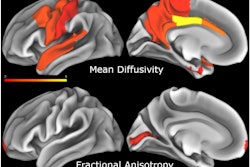
 A graphical abstract of the study. Image courtesy of RSNA.
The primary analysis revealed no evidence of a difference in the ratio of lactate to Glx between participants with Alzheimer’s disease and healthy participants across all regions of interest, the authors reported.
However, an exploratory analyses revealed that participants with Alzheimer’s disease had reduced signals for medial temporal lactate (0.7 vs. 0.5, p = 0.04) and Glx (0.5 vs. 0.48, p = 0.03) compared with controls. Finally, a strong correlation (r = 0.73) was observed between DMI and FDG PET, the authors noted.
“Deuterium metabolic imaging was highly aligned with fluorodeoxyglucose [FDG] PET in participants with Alzheimer disease (AD) but was unable to help differentiate participants with AD from age-matched controls,” the group wrote.
In a related study also in the April 8 issue Radiology, researchers at Yale University in New Haven, CT, appear to have developed a protocol that could help with clinical use of DMI.
The researchers designed a parallel acquisition strategy that obtains metabolic DMI data during a comprehensive, multicontrast MRI (fluid-attenuated inversion recovery [FLAIR] and T1-, T2-, and susceptibility-weighted) without adding scanning time.
“Parallel MRI scan acquisition technology was a practical solution to obtain both high-quality anatomic and metabolic scans without prolonging the scanning duration compared with an MRI-only protocol,” noted first author Yanning Liu, MD, and colleagues.
The authors used a dual-tuned proton-deuteron head coil to develop the parallel sequence and evaluated it in five healthy volunteers and one patient with a grade 4 brain tumor. Two healthy volunteers and the patient drank a solution of 65 grams of deuterated glucose dissolved in water and underwent imaging between 60 and 120 minutes later.
The technique worked, with reasonable quality clinical and glucose DMI scans acquired in 42 minutes, the authors noted.
“MRI scan quality, DMI sensitivity, and information content are preserved in the parallel MRI DMI acquisition method,” the group wrote.
In an editorial accompanying the studies, John Port, MD, PhD, of the Mayo Clinic in Rochester, MN, noted that while the studies lack large numbers of participants and extensive results, they are important because they show the feasibility of the glucose DMI technique and its clinical diagnostic potential.
The authors have overcome numerous technical limitations to create a vision of the future of human metabolic imaging, Port wrote.
“As we have done in the past, improvements in MRI technology through clever engineering will pave the way for an entirely new kind of imaging, perhaps transforming medicine yet again,” he concluded.


.fFmgij6Hin.png?auto=compress%2Cformat&fit=crop&h=100&q=70&w=100)


.fFmgij6Hin.png?auto=compress%2Cformat&fit=crop&h=167&q=70&w=250)


