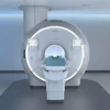
MRI-targeted biopsies performed inside MRI scanners (in-bore) are effective at finding significant disease in patients with suspected prostate cancer, according to a study published July 29 in Radiology.
The result is from an analysis of 780 procedures performed over eight years at the European Institute of Oncology in Milan, Italy, and is particularly relevant for patients who may have early stages of the disease, noted lead author Caterina Sattin, MD, of the University of Milan, and colleagues.
"Given the high risk of undergrading [grade group] 1 cancers, it is important to integrate MRI findings and clinical metadata with in-bore MRI-targeted biopsy results to personalize treatment decisions and avoid potential undertreatment of patients with prostate cancer," the investigators wrote.
In-bore MRI-targeted biopsy is a highly effective method for grading the severity of prostate cancer, as it enables precise needle placement. Yet whether the cancer grading from the in-bore biopsy matches the final grading determined after surgery, and what factors affect how well these grades match, is underexplored, the authors explained.
To that end, they evaluated factors affecting the detection rate of prostate cancer in patients who underwent in-bore MRI-targeted biopsy and their influence on histologic concordance between biopsy grade group and surgical grade group.
According to the analysis, detection rates for grade group (GG) 2 or higher prostate cancer depended on the size of the MRI target lesion and the Prostate Imaging Reporting and Data System, or PI-RADS, category. Concordance between biopsy and surgical GGs was moderate (60.2%), with a risk of undergrading, the researchers reported.
Key results included the following:
In-bore MRI-targeted prostate biopsy detection rates were higher for larger lesions (> 10 mm and PI-RADS category ≥ 4).
The concordance between in-bore biopsy and surgical GG was higher when biopsy grades were GG 2 or 3 but lower when they were GG 1, 4, or 5.
Differences between biopsy and surgical grades did not depend on lesion size or PI-RADS category.
Further, there was no significant difference in performance whether the in-bore MRI-targeted prostate biopsy procedure was performed manually or by robotic biopsy systems, the authors noted.
"The concordance between the biopsy grade groups and surgical grade groups was moderate to substantial overall, but varied by [biopsy grade group], with a high risk of undergrading when GG 1 cancers were found," the group concluded.
Ultimately, downgrading and upgrading GG during MRI-targeted biopsies can have important management consequences related to potential undertreatment or overtreatment, the authors concluded.
In an accompanying editorial, Laurent Milot, MD, PhD, of the University of Lyon in France, wrote that the study reveals both the diagnostic strengths and limitations of in-bore MRI-targeted biopsy, which is crucial as the field continues its trajectory toward personalized, minimally invasive care.
"[The study] reiterates a central truth in oncologic imaging: Precision in localization does not equate to perfection in characterization," Milot wrote. "The ability to accurately guide a needle to a lesion is only one part of the diagnostic equation. The biologic behavior of prostate cancer, its architectural complexity, and its multifocal nature make it inherently elusive. This is why risk-adapted strategies that combine imaging, targeted histologic analysis, prostate-specific antigen kinetics, and patient-specific factors (including the preferences of the patient) are not just helpful – they are essential."
The full study is available here.