
Researchers using fMRI have identified a brain region associated with why people sometimes fail to apply their own moral standards to their own behavior, according to a study published March 19 in Cell Reports.
A team led by senior author Hongwen Song, PhD, of the University of Science and Technology of China in Hefei, found that the ventromedial prefrontal cortex (vmPFC) showed less activity and reduced connectivity to other decision-making brain regions in participants who judged dishonest behavior harshly in others but rated their own similar behavior more leniently. In participants deemed "morally consistent," vmPFC activation was similar during both behavioral and judgment tasks.
To test whether the vmPFC plays a causal role, the investigators stimulated participants' vmPFCs using transcranial temporal interference stimulation, a noninvasive method, prior to the tasks. Stimulated participants showed higher levels of moral inconsistency compared with those who received mock stimulation, they said.
"Our findings suggest that we should treat moral consistency like a skill that can be strengthened through deliberate decision-making," Song said in a statement released by the journal. "These findings have huge implications for education and AI."
Access the full study here.


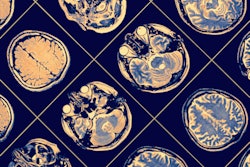


![Overview of the study design. (A) The fully automated deep learning framework was developed to estimate body composition (BC) (defined as subcutaneous adipose tissue [SAT] in liters; visceral adipose tissue [VAT] in liters; skeletal muscle [SM] in liters; SM fat fraction [SMFF] as a percentage; and intramuscular adipose tissue [IMAT] in deciliters) from MRI. The fully automated framework comprised one model (model 1) to quantify different BC measures (SAT, VAT, SM, SMFF, and IMAT) as three-dimensional (3D) measures from whole-body MRI scans. The second model (model 2) was trained to identify standardized anatomic landmarks along the craniocaudal body axis (z coordinate field), which allowed for subdividing the whole-body measures into different subregions typically examined on clinical routine MRI scans (chest, abdomen, and pelvis). (B) BC was quantified from whole-body MRI in over 66,000 individuals from two large population-based cohort studies, the UK Biobank (UKB) (36,317 individuals) and the German National Cohort (NAKO) (30,291 individuals). Bar graphs show age distribution by sex and cohort. BMI = body mass index. (C) After the performance assessment of the fully automated framework, the change in BC measures, distributions, and profiles across age decades were investigated. Age-, sex-, and height-adjusted body composition reference curves were calculated and made publicly available in a web-based z-score calculator (https://circ-ml.github.io).](https://img.auntminnie.com/mindful/smg/workspaces/default/uploads/2026/05/body-comp.XgAjTfPj1W.jpg?auto=format%2Ccompress&dpr=2&fit=crop&h=167&q=70&w=250)


