The Developing Fetus: First and Second Trimesters CD-ROM or video sets
American Institute of Ultrasound in Medicine, Laurel, MD, 2003, $495 for AIUM members; $990 for nonmembers
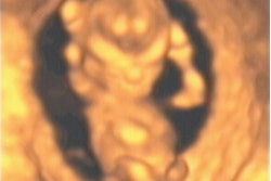
Ultrasound continues to rapidly provide answers to important questions regarding fetal health, thanks to advances in technology and clinical research. The Developing Fetus: First and Second Trimesters is a set of eight CDs, or VHS tapes, consisting of sixteen lectures that were delivered in Montreal at an AIUM meeting.
The courses are excellent in the breadth and depth of coverage, and they are delivered by twelve prominent lecturers. The entire set provides complete sonographic coverage for evaluating the fetus, from the earliest detection of the gestational sac to early anatomic survey and risk assessment for trisomies.
Many lectures are anatomically-based, with separate talks covering the fetal ventricle, spine, face, thorax, GI tract, GU tract, skeletal conditions, and heart. Abnormal findings are covered in presentations on failing IUP, ectopic pregnancies, and borderline findings in the fetus. Additional lectures cover sonoembryology, the early anatomic survey, the genetic sonogram, and the discussion of risk with patients.
Most lectures present information at an introductory level before building up to more advanced topics. The spiral-bound syllabus that comes with the set contains notes, figures, and references for many of the presentations. Up to 10 CME credits are awarded by the AIUM for completion of the program.
Dr. Peter Doubilet, Ph.D., offers a particularly outstanding talk on sonographic markers at 15-20 weeks gestation. He first defines the sonographic criteria for markers, such as thick nuchal cord and echogenic bowel, then offers plenty of examples. He wraps up his lecture by applying the results to patients with different, pre-scan risks of Down syndrome.
All of the speakers give practical advice on effective communication of sonographic findings, and the associated good or bad risk to parents. This issue is important as physicians must juggle abnormal findings and their meanings while not generating undue anxiety for the parents.
This program did have several weaknesses. The most obvious one is that of resolution: This is essentially a home movie (352 x 234 pixels) of a high resolution PowerPoint presentation (probably 1,024 x 768 pixels). Often, the particular feature being discussed is quite difficult to see on the monitor -- particularly if more than one image is being projected on the screen.
Secondly, there are vast differences in the quality of accompanying notes and figures. Some lecturers included copies of all of their slides; others included important tables and figures. Yet others only submitted a paragraph of information and neglected references altogether. In one extreme case, the person who gave the lecture and the person who wrote notes in the syllabus were entirely different.
Finally, the lecture on skeletal anomalies is not all that relevant to most ultrasound practices. Although beautifully executed with outstanding animation, pretty pictures, and clever diagrams, the practice of identifying a subtle skeletal finding, searching the OMIM (Online Mendelian Inheritance in Man) database for associated syndromes, and presenting this list to the referring physician may not be a useful exercise in the long-run.
Overall The Developing Fetus First and Second Trimesters effectively blends state-of-the art obstetric ultrasound with current clinical research and practical obstetric patient management.
By Dr. Danny DonovanAuntMinnie.com contributing writer
March 16, 2004
Dr. Donovan is a third year resident at Methodist University Hospital in Memphis, TN. He is interested in pursuing a fellowship in cardiac and noninvasive vascular imaging.
The opinions expressed in this review are those of the author, and do not necessarily reflect the views of AuntMinnie.com.
Copyright © 2004 AuntMinnie.com


