Dear Ultrasound Insider,
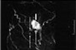
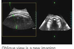
Two-dimensional sonography views of the uterus and endometrium have been typically confined to sagittal and transverse images. But with 3D, they can now be imaged in the coronal plane.
And 3D reconstructed images of the endometrium in the coronal plane can provide additional findings in 30% of routinely performed transvaginal pelvic sonography exams, and in half of the cases that are abnormal on 2D imaging, according to researchers from Vanderbilt University Medical Center in Nashville, TN.
This month's Ultrasound Insider Exclusive story on the Vanderbilt research is part of our continuing coverage from the annual meeting of the American Institute of Ultrasound in Medicine (AIUM) in March.
As an Ultrasound Insider subscriber, you have access to this story before it is published for the rest of our AuntMinnie.com members. To read more about the added value of 3D, click here.
Do you have an idea for a topic you'd like to see covered? As always, please feel free to drop me a line.