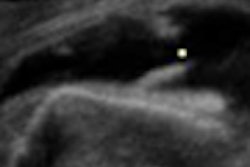
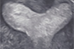
Real-time 3D contrast-enhanced transcranial ultrasound can help visualize major cerebral vascular vessels, according to research published online in Ultrasound in Medicine and Biology.
"These preliminary results suggest that [real-time 3D contrast-enhanced] transcranial US and [real-time 3D contrast-enhanced] transcranial US with phase aberration correction have the potential to greatly impact the field of neurosonology," wrote a research team led by Nikolas Ivancevich from Duke University in Durham, NC.
The Duke researchers scanned 17 healthy volunteers using a 3D ultrasound scanner (Volumetrics Medical Imaging, Durham, NC) and the Definity microbubble contrast agent (Lantheus Medical Imaging, North Billerica, MA). Exclusion criteria for the study included pregnancy or lactation, history of neurologic disease, congenital heart defect, severe liver dysfunction, respiratory distress syndrome, cardiac shunts, and hypersensitivity to octafluoropropane (Ultrasound in Medicine and Biology, April 18, 2008).
Seventeen of the patients were scanned via a single temporal window, while nine were also scanned via the suboccipital window. A vascular sonographer and a neurologist then independently reviewed the temporal and suboccipital volumes from each patient using the scanner's orthogonal 3D display as well as the offline 3D Doppler volume renderings. Both reviewers were asked to note the presence or absence of major cerebral vessels.
For 71% of the subjects, both observers identified the ipsilateral circle of Willis from the temporal window. In addition, the entire circle of Willis was visible in 59% of the cases, according to the researchers. In the suboccipital window, both reviewers detected the entire vertebrobasilar circulation in 22% of the subjects and the basilar artery in 44%.
"After performing phase aberration correction on one subject, we were able to increase the diagnostic value of the scan, detecting a vessel not present in the uncorrected scan," the authors wrote.
Future Duke research will explore the sensitivity and specificity of real-time 3D contrast-enhanced transcranial ultrasound using digital subtraction angiography or MR angiography as a gold standard. The Duke researchers said they will also perform phase aberration correction on more subjects to further determine its added value for transcranial ultrasound.
"We are also pursuing advances in phase aberration correction to increase the diagnostic value of this technique, especially in conjunction with moving target techniques for echoes from flowing contrast agent," they wrote.
By Erik L. Ridley
AuntMinnie.com staff writer
June 13, 2008
Related Reading
Microbubbles speed ultrasound-enhanced clot lysis in stroke, February 14, 2006
Ultrasound results suggest possible etiology for restless legs syndrome, November 21, 2005
Transcranial ultrasound of little use as stroke therapy, January 24, 2005
Copyright © 2008 AuntMinnie.com


