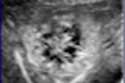
Breast ultrasound computer-aided detection (CAD) appears to help doctors more consistently find lesions smaller than 1 cm, according to a study conducted by Chinese researchers. And using a single reader with CAD produces comparable results to using two readers to interpret breast ultrasound, the group found.
A presentation at the 2008 annual conference of the National Consortium of Breast Centers (NCBC) described the research from the People's Liberation Army General Hospital in Beijing, China, evaluating the use of CAD with breast ultrasound in diagnosing breast cancer. The study results also are to be published this month by the American Journal of Clinical Oncology.
From November 2005 until January 2007, Dr. Junlai Li and colleagues reviewed 100 breast ultrasound scans from 99 patients, which were taken using scanners from a number of manufacturers: Sequoia 512 from Siemens Healthcare of Erlangen, Germany; Logiq 7 and Logiq 9 from GE Healthcare of Chalfont St. Giles, U.K.; HDI 5000 and iU22 from Philips Healthcare of Andover, MA; and Aplio XG from Toshiba Medical Systems of Tokyo. The team used B-CAD version 2 software from Medipattern of Toronto.
They found 64 malignant breast lesions and 36 benign breast lesions; all 100 diagnoses were confirmed by pathology through surgery or coarse-needle aspiration biopsy. Cases were read by a single physician without the use of CAD, a single physician using CAD, and two physicians performing double reading without CAD.
The team found that for lesions greater than 1 cm, there was no difference in diagnostic accuracy between the three reading techniques. Single doctors reading ultrasound studies with the help of CAD had a diagnostic accuracy of 100%, finding all 64 of the malignant breast lesions in the study cohort; single doctors reading scans without the use of CAD found 60 out of 64 malignancies, for an accuracy rate of 93%; and the two doctors reading together found 63 out of 64 cancers, for an accuracy rate of 98%.
CAD was able to increase diagnostic accuracy in identifying lesions less than 1 cm, the study found. Accuracy increased by 44% when single readers using CAD and single readers not using the technology were compared.
The difference in diagnostic accuracy between a single reader using CAD and double readers was not significant, the team wrote, suggesting that imaging centers could have room to decide which of these techniques is most cost-effective without fear that diagnostic accuracy would be lost.
But even with its high sensitivity, CAD isn't a substitute for good, old-fashioned experience, according to Li.
"Doctors should analyze ultrasonic images of breast lesions assisted by CAD, then judge the suspicious sites CAD identifies and make the diagnostic decision using their own clinical experience," Li wrote.
By Kate Madden Yee
AuntMinnie.com staff writer
October 10, 2008
Related Reading
Breast ultrasound CAD performance varies in ethnic populations, September 5, 2008
Breast CAD improves classification accuracy, cuts ultrasound biopsies, February 29, 2008
Copyright © 2008 AuntMinnie.com


