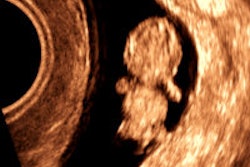
AuntMinnie.com presents the 20th in a series of columns on the practice of ultrasound from Dr. Jason Birnholz, one of the pioneers of the modality.
The RSNA meeting is emblematic of the holiday season. There is the congregation of friends and colleagues from all over. There is the sharing of knowledge and experience, and there are some fabulous parties. The exhibit halls showcase Santa's newest technoproducts.
Manufacturers tend to aim for RSNA specifically for new releases, anticipating first-quarter product launches. I spend a lot of time booth hopping, which I think of as educational, along with the many courses.
White papers and an old friend
One booth I visited was Zonare Medical Systems, where I inquired specifically about the company's 20-MHz transducer. I was handed a white paper titled "Two-way continuous transmit and receive focusing in ultrasound imaging"; it identified the core feature of Zone sonography. There were several "ideal" beam plots with transmit, receive, and dual-focusing options.
 Dr. Jason Birnholz.
This was a "white paper," self-published in-house. White papers resemble peer-reviewed articles visually, but the content typically lacks analytic or comparative material. They are usually well-written and very educational, but they are editorials or marketing pieces. I also like to see clinical results for anything new so I can decide for myself if this will extend my practice in a meaningful way.
White papers often have an outside consultant as lead author, with two to four contributors from within the company. For this paper, I was really happy to see that the first author was Thomas Jedrzejewicz, identified as a medical ultrasound consultant in Austin, TX. I have known Tom for at least 30 years, although I regret I haven't seen him lately.
Tom is the epitome of a scholar and gentleman. He was with Acuson for a long time, and he had a lot to do with the initiation of energy Doppler. Tom is on the engineering side of ultrasound, so a lot of clinical users will probably not know how instrumental he has been in the development of modern ultrasound imaging technology. One of my intentions for this coming year is to feature some outstanding ultrasound people, like Tom, who have contributed greatly (and often unobtrusively) to our field.
Tom had a lot to do with the development of 2D energy Doppler. I prefer "energy" Doppler to the much more common usage of "power" Doppler. Doppler measures the frequency shift of a beam intersecting a slower-moving target. Beam and target velocities are directionally additive. The clinical utility of Doppler is enhanced by using the square of the frequency shift, a value that is always positive. Kinetic energy is 0.5 x mass x velocity squared. The squared velocity is the common factor, hence the use of "energy." Power refers to the rate that work is done (or that energy is utilized), so it seems to me that the catchy-sounding power Doppler is a misnomer.
This pickiness may be almost as pointless as arguing about endo- (where the probe is) versus trans- (where the beam goes) vaginal ultrasound. Actually, it would be double transvaginal, since you've got sending and receiving for each conventional ultrasound image.
A lot of people would say that common usage for long enough validates a term; however, I think this kind of nomenclatural sloppiness is one of the things that continue to marginalize ultrasound as a serious discipline.
Jellyfish
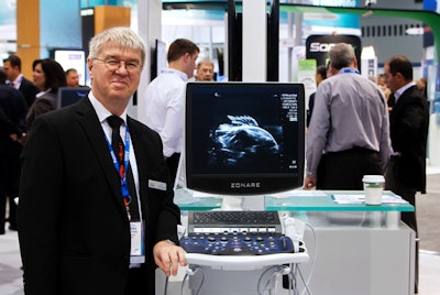
 Ludwig Steffgen from Zonare. Image courtesy of Dr. Jason Birnholz.
The smiling figure with the proprietary demeanor is Ludwig Steffgen, Zonare's director of marketing for premium products, with a frozen frame of a tropical fish acquired with Zonare's 14-MHz probe. Ludwig told me that he learned a lot of anatomy while working as a pathology technician before he started teaching. He did the ultrasound technologist training program at Stanford when he worked for Acuson. We share a lot of interests, like photography and ultrasounding oddball targets. Ludwig shared with me several video clips of test objects, one of which was of a tiny freshwater jellyfish made with the 20-MHz transducer.
Video courtesy of Ludwig Steffgen, Zonare.
I pretty sure Ludwig mentioned that he had discovered this rare kind of jellyfish cave diving. Both the adventurousness and image quality impressed me. There really isn't any background noise at all, usually a big problem at higher frequencies. Detail is great, motion is tracked smoothly, and the particulates recirculating in the water bath are distinct.
Perils and problems of new probes
I would really have liked to see some clinical images, even if they were of the eye or skin via a water path, in case attenuation might be an issue with this first version of the probe. One of the thoughts I had was to reconfigure the transducer at about a 30° angle on a long handle as an endovaginal probe (or at right angles for the cervix and prostate). The transvaginal portal (since we're now talking about the beam path) would minimize attenuation and multiple reverberation problems, and I would anticipate that contrast and spatial resolution features should make identification of endometriosis and adhesions very certain, should result in enhanced evaluation of early first-trimester embryos, and might provide a means of finding precancerous changes in the ovary, which we do not have in any imaging technique yet.
As much as I would like to test these surmises myself right now, equipment development has become a very difficult and precarious proposition for manufacturers. The direction of development has to be initiated by engineering, which is constrained by physical laws that have to do with wave generation, propagation, and reception, as well as by regulatory laws that concern energy transfer and safety issues. There are also constraints of costs of materials and manufacturing. One of the consequences is that the business sides of companies frequently aim at improving existing applications rather than speculating in brand new applications without demonstrated clinical utility.
There is another issue that has to do with transducers themselves. I am intending to have an article devoted to transducers soon. Modern transducers are multielement arrays, sometimes linear, sometimes 2D with off-axis elements. There are new materials for the individual elements and designs to limit cross-talk, a major problem area.
The transducer array needs to be tuned exactly to the ultrasound system, which is a hardware issue, and then there is integration with the entire chain of software modules. So far, from just ordering up a transducer and popping it into a system, every little and every big change requires a lot of system-wide work. You all understand this from thinking about diseases and bodies; most of the time, you have to think systemically.
From cooperative to commercial
Most of the time, we have the luxury of using equipment without knowing what manufacturers have had to go through to get a reliable workhorse unit out to the public globally. Once upon a time, radiologists and x-ray imaging and ancillary device manufacturers had a special relationship that revolved around research. Government and foundation grants were the main source of support for our colleagues doing bench research, while clinical imaging research was more often than not supported by equipment manufacturers.
Typically, manufacturers provided equipment without charge for onsite use, but they did not participate in the research project, which was designed, conducted, analyzed, and reported by the researchers independently. The researchers were salaried by their institutions, not paid by the manufacturers, and studies were performed for free whether they were on volunteers or patients fitting the study protocol. The institutions did not charge any "overhead" to the manufacturers, and they contributed space and whatever supplies, ancillary equipment, or correlative lab or pathology services were required.
The institutions encouraged such liaisons. Research was a voluntary part of a participating radiologist's responsibilities, melded with one's daily clinical responsibilities. I believe this was the common practice here and throughout Europe and Asia.
Those really were the good old days. Most of the work was with plain radiography, x-ray fluoroscopy, or radionuclide imaging. There were no institutional review boards (IRBs), although a radiologist always functions as a radiation safety officer, and actual imaging procedures were always conducted under the same stringent rules that pertain to requested studies. IRBs are a very good thing, though research prior to their inception was scientific and done with regard to patient welfare. Voluntary collaborative research accounted for a lot of clinical advances over all types of medical imaging in the past. It works because both sides are extremely well-educated and both want sincerely to achieve better patient care.
I have been very fortunate to have had the opportunity to collaborate with a lot of the ultrasound manufacturers over the past 35 years. The best times were interactions with engineers. I was always impressed by their willingness to embrace novelty and to learn about clinical realities, and I learned an enormous amount about the foundations of ultrasound imaging from them. Most of all, getting a chance to work with the latest equipment enabled me to do better for patients referred to me. I believe that all of you would feel the same if you were asked to contribute in this same way.
A suggestion for manufacturers for 2015
What I have been saying is that direct collaboration between clinical users and equipment manufacturers is a win-win situation. In the past, most of the collaborations were centered on academic institutions, which is a centralized process. I want to suggest something distributive involving private-practice ultrasound facilities. When manufacturers have something they think may be ready for prime time, they should enlist cooperatively as few or as many facilities as they can that are already using their equipment.
If the improvement already has U.S. Food and Drug Administration (FDA) clearance, then no special research or test allowance is necessary, and there are independent IRBs for new applications. Companies should provide equipment, transducers, software, or ancillary devices for free during the test period, and facilities should solicit patients or volunteers for free exams according to the study intent and protocol. All collaborating facilities should have the chance to pool their findings, and all findings should be available generally whenever they appear to have scientific consequence.
Just think about all the clinical exams done in the world every day. If only we could find a way to share our findings and experiences effectively, clinical sophistication and quality control would race throughout our community. If manufacturers only asked us to collaborate with them routinely, imagine the consequences for our immediate technical progress. It makes a really good wish for the coming year.
Dr. Jason Birnholz was one of the few advanced academic fellows of the James Picker Foundation, and he has been a professor of radiology and obstetrics. He is a fellow of the American College of Radiology and the Royal College of Radiology, and he was an associate fellow of the American College of Obstetricians and Gynecologists.
The comments and observations expressed herein do not necessarily reflect the opinions of AuntMinnie.com.


