In what they are calling a major advance, researchers from Michigan have created a 3D model of a human heart using data from two separate cardiac imaging techniques: CT and echocardiography. They believe that such hybrid 3D models will be more accurate than those created from just one imaging modality.
The study team from Spectrum Health Helen DeVos Children's Hospital in Grand Rapids, MI, hailed the proof-of-concept study as the first use of hybrid imaging in the creation of a 3D heart.
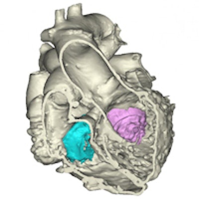
 3D image of heart model. Image courtesy of Spectrum Health.
Hybrid 3D printing integrates the best aspects of two or more imaging modalities, potentially enhancing diagnosis and improving interventional and surgical planning, said lead author Jordan Gosnell, a cardiac sonographer at the hospital. Previous 3D printing models used only a single modality, which is less accurate than merging two or more datasets.
The study also opens the way for hybrid 3D printing techniques to be used in combination with a third modality: cardiac MR, the study team said in a statement accompanying the results.
First, the researchers used software to register images from CT and 3D transesophageal echocardiography (TEE) scans; they then selectively integrated the datasets to produce the anatomic model of the heart. The results provide more detailed and anatomically accurate 3D renderings and printed models than are available from a single modality, which may allow clinicians to improve their diagnosis and treatment of heart disease.
Each imaging modality has different strengths, and combining the modalities leads to improved results, according to the researchers:
- CT enhances the outside anatomy of the heart.
- MRI is superior for the interior of the heart, including the right and left ventricles and the heart's muscular tissue.
- 3D TEE offers the best visualization of valve anatomy.
The work was presented at the 2015 Catheter Interventions in Congenital, Structural, and Valvular Heart Disease (CSI) meeting in Frankfurt, Germany, by study co-author Dr. Joseph Vettukattil, who has performed research with 3D and 4D echocardiography. Vettukattil developed the use of multiplanar reformatting (MPR) in echocardiography to evaluate complex heart defects.
"This is a huge leap for individualized medicine in cardiology and congenital heart disease," Vettukattil said in the statement. "The technology could be beneficial to cardiologists and surgeons. The model will promote better diagnostic capability and improved interventional and surgical planning, which will help determine whether a condition can be treated via transcatheter route or if it requires surgery."