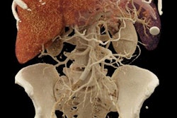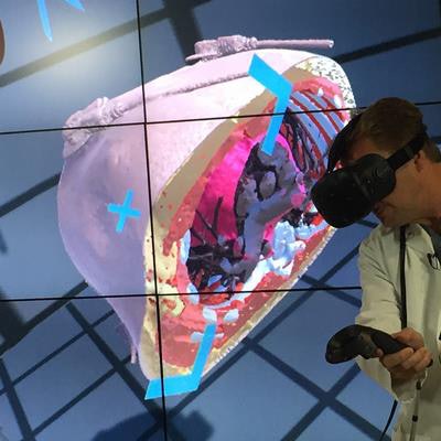
These are still the early days for advanced visualization and 3D printing, but you'd hardly know it from the operation at Jump Trading Simulation and Education Center. Researchers are hard at work building a library of 3D heart models designed to realize the potential of 3D modeling for education, surgical planning, and other applications.
At its center in Peoria, IL, the researchers manipulate wall-sized 3D displays and work with engineers and cardiologists who are dedicated to expanding the boundaries of 3D simulation in medicine. Jump is building a library of peer-reviewed 3D heart models created using data from actual imaging scans, with the goal of using simulation tools like 3D printing to improve healthcare quality.
"Just as the airline industry went through a time when they began simulation to improve quality, healthcare is going through that right now," said Dr. Matthew Bramlet, pediatric cardiologist and lead researcher for Jump's Advanced Imaging and Modeling (AIM) project, in an interview with AuntMinnie.com.
Human and financial resources
Jump's signature project is the development of a 3D heart library in cooperation with the U.S. National Institutes of Health (NIH). It has some major backing to accomplish this and other goals: The center is collaboration between the Catholic nonprofit organization OSF HealthCare and the University of Illinois College of Medicine, and it was bolstered last month by a new $10 million endowment from William Shepard, who added it to his original $5 million investment based on the center's early successes.
The Jump center funds engineers who develop 3D visualization software to create what it calls "printable 3D hearts" for the benefit of patients, providers, and the NIH. The models are delivered to the NIH's 3D Print Exchange Heart Library and will permit further expansion of the Advanced Imaging and Modeling project, designed to educate patients and their families through advanced imaging, virtual augmented reality, and 3D models. So far, the center has contributed about 60 hearts to the NIH database.
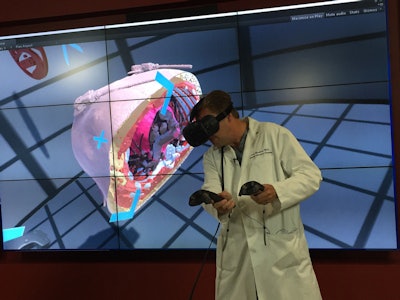 Exploring anatomy with the HTC Vive simulator at Jump. Image courtesy of Dr. Matthew Bramlet.
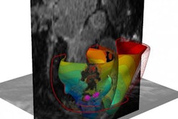
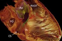
"We're taking standard medical images, MRI and CT primarily, and translating them into 3D models that can be displayed either in a printed form or in virtual reality," Bramlet said. "It's our ability to integrate this into health systems that has an impact on patient care."
If a surgeon has a patient with a particular type of pathology, he or she can order a 3D-printed heart from the NIH library, and the heart is then shipped for surgical planning. Or, a medical student might want to learn about the variations of tetralogy of fallot, a complex 3D problem that can be challenging to teach by textbook.
"If I can print a model out that they can base their learning on, then that's going to improve their understanding, improve their retention, and hopefully improve their medical decision-making related to the physiology as related to anatomy," Bramlet said.
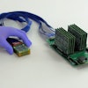
The center uses a Vive (HTC) virtual reality system to interact with MR and CT images of anatomy. Caterpillar 3D printers and Tango flexible printing materials are used to create malleable 3D hearts. Current software projects include a web-based reviewing tool that overlays 3D content onto 2D data, Bramlet said.
Peer review for images
Jump believes that its models -- based on real patient data -- are an improvement over other renditions that have been converted into models. By creating a quality metric for image data, the Jump team can effectively peer-review models and confer a stamp of approval on which people can base their medical education, Bramlet said.
Another purpose of the $10 million endowment is to provide a 3D-printed heart to any clinical pediatric cardiologist or surgeon who needs one, at no cost to the patient, he said.
What he is sure of is that doctors exposed to 3D models are forever changed by them.
"Not everybody has been impacted by the 3D model yet, but if you were to interview surgeons around the nation who have held a 3D heart in their hand, they'll say they won't operate without it," Bramlet said. "Every time we print a 3D heart, I learn something new about that heart. Does what I learn change or impact the surgery or surgery planning? In the majority of cases it does not, but it has in a small number of cases, and so as the complexity increases, so does the value of a 3D-printed model."
The wide-open nature of the NIH data makes its potential quite broad, he said. The NIH focuses on housing the database, which includes not only anatomical data but also biologic proteins, viruses, and biomedical content.
"My favorite story is from a researcher who held an influenza virus in his hand for the first time, and within 15 minutes knew how to lay down a protein over it to create a vaccine that he had not appreciated without the 3D model in front of him," Bramlet said. "But in order for him to know that, he had to make sure the 3D model was accurate. We're working with experts in cardiology to come up with a toolset that allows us to put that stamp of approval."
A YouTube video showing 3D simulation at Jump can be seen here.

