While detecting subclinical atherosclerosis is useful, quantifying the plaque burden is even more valuable. A 3D vascular ultrasound method can reliably perform this task and help to estimate cardiovascular risk, according to research published in the July 18 issue of the Journal of the American College of Cardiology.
Among nearly 4,000 patients in the Progression of Early Subclinical Atherosclerosis (PESA) study, 3D vascular ultrasound was a feasible and reproducible method for quantifying early carotid and femoral atherosclerotic burden, a Spanish research team found. Quantitative plaque burden -- particularly in the femoral arteries -- was associated with conventional cardiovascular risk factors. What's more, the method more closely reflected estimated cardiovascular risk, compared with just the presence of plaque alone.
"This inexpensive and radiation-free technology thus has the potential to become a key large-scale screening tool for identifying at-risk individuals," wrote the team led by Dr. Beatriz López-Melgar, PhD, of Centro Nacional de Investigaciones Cardiovasculares Carlos III (CNIC) in Madrid.
Improving risk stratification
Evidence is increasing to reinforce the value of detecting atherosclerotic lesions for cardiovascular risk assessment. Although 2D vascular ultrasound is well-suited for detecting and grading the extent of atherosclerosis in different arterial territories, methods for quantifying plaque burden with 2D vascular ultrasound have differed between studies, and there is no consensus on which measurement should be used or which territory is best for predicting cardiovascular risk, according to the team.
A semiautomated 3D vascular ultrasound method has been proposed for quantifying peripheral atherosclerotic burden, but the method hasn't yet been evaluated in large population studies. In addition, the association of plaque volume with cardiovascular risk factors has not been explored, the authors wrote.
As a result, they set out to evaluate the feasibility and reproducibility of the method and to provide reference values of carotid and femoral plaque burden distribution by 3D vascular ultrasound in asymptomatic middle-aged individuals. They also wanted to assess the relationship between plaque burden and cardiovascular risk factors, as well as explore the potential added value of plaque volume quantification over plaque detection alone (JACC, July 18, 2017, Vol. 70:3, pp. 301-313).
PESA study
The research included 3,860 (92.2%) participants in PESA, an ongoing observational prospective cohort study involving 4,184 employees of Banco de Santander in Madrid. The project aims to characterize early subclinical atherosclerosis burden and the determinants of atherosclerosis presence and progression using different noninvasive techniques in volunteers without prior cardiovascular disease. The volunteers were enrolled in the study between June 2010 and February 2014. The participants, 63% of whom were men, had an average of 45.8 years.
At their baseline visit, the participants received a 3D vascular ultrasound study, clinical interviews, a standardized lifestyle questionnaire, a physical exam, a fasting blood draw, urine sample collection, and a 12-lead electrocardiogram. Their American College of Cardiology/American Heart Association atherosclerotic cardiovascular disease score was quantified and classified according to 10-year risk as low (< 5%), intermediate (5% to 7.5%), or high (> 7.5%). The participants were prospectively followed with repeat visits at three and six years.
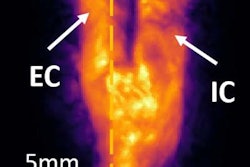
Using an iU22 ultrasound scanner (Philips Healthcare) with a VL 13-5 3D volume-linear array transducer, 3D vascular ultrasound was performed in a standardized fashion on bilateral carotid and femoral arteries to determine the number of plaques and affected arterial territories. The images were analyzed offline at the CNIC Imaging Core Laboratory using version 10.2 of the Qlab software (Philips). Global plaque burden was then quantified as the sum of all plaque volumes. The researchers used linear regression analysis and proportional odds models to evaluate the associations of plaque burden with cardiovascular risk factors and estimated 10-year cardiovascular risk.
Higher plaque burden in men
In agreement with previous studies using 2D vascular ultrasound, men had more than double the global plaque burden compared to women:
- Median global plaque burden in men: 63.4 mm3 (interquartile range [IQR]: 23.8 to 144.8 mm3)
- Median global plaque burden in women: 25.7 mm3 (IQR: 11.5 to 61.6 mm3)
The difference was statistically significant (p < 0.001). In addition, plaque burden was higher in the femoral territory:
- Median plaque burden in femoral territory: 64 mm3 (IQR: 27.6 to 140.5 mm3)
- Median plaque burden in carotid territory: 23.1 mm3 (IQR: 9.9 to 48.7 mm3)
The difference was also statistically significant (p < 0.001). In other findings, plaque burden increased significantly with age (p < 0.001):
- Median plaque burden for ages 40-44: 31.2 mm3 (IQR: 18.7 to 121.5 mm3)
- Median plaque burden for ages 45-49: 55 mm3 (IQR: 19.7 to 121.2 mm3)
- Median plaque burden for ages 50-54: 78.9 mm3 (IQR: 27.7 to 176.6 mm3)
Femoral plaque burden
Delving further into the data, the researchers found that age, sex, smoking, and dyslipidemia were also more strongly associated with femoral rather than carotid disease burden. However, hypertension and diabetes showed no territorial differences, according to the group.
There was a "higher plaque volume in the femoral territory in both sexes, whereas plaque presence was more frequent in the femoral arteries of men but in the carotid arteries in women," the authors wrote. "Furthermore, age-related changes in plaque volume tended to be more rapid in the femoral arteries of men. Thus, compared with plaque detection, plaque volume quantification unveils novel sex- and artery-related differences in the early development of atherosclerotic disease."
Also, plaque volume showed a significant correlation with higher cardiovascular risk category, independent of the number of plaques or territories affected (p < 0.001).
"Global plaque volume, by integrating plaque presence, number, and plaque size, is a more comprehensive index of disease burden that may better reflect individual susceptibility, and thus partially explain the mismatch between risk profile and plaque presence," the authors wrote.
An important step forward
The research marks an important step forward in the measurement of atherosclerotic burden, making it easier to implement not only epidemiological studies and studies of new therapies, but also the strategy of "treating arteries," said Dr. J. David Spence in an accompanying editorial.
"Measurement of carotid plaque burden has come a long way since 1990; this study promises to bring it to the mainstream of cardiovascular medicine," wrote Spence, a professor at Western University in London, Ontario, Canada.


