A newly developed contrast-enhanced ultrasound (CEUS) Vesical Imaging-Reporting and Data System (VI-RADS) helps both novice and expert readers to evaluate muscle-invasive bladder cancer (MIBC), researchers reported September 10 in Radiology.
"Contrast-enhanced ultrasound (CEUS) with microbubbles makes it possible to clearly identify and assess the three-layer structure of bladder wall by means of real-time perfusion imaging," noted a team led by Jing Han, MD, of the Sun Yat-sen University Cancer Center in Guangzhou, China, which introduced the CEUS VI-RADS system.
While CEUS is not used clinically for this indication as commonly as multiparametric MRI (mpMRI), it offers an opportunity to evaluate muscle invasion preoperatively. Han and colleagues explained that mpMRI may not be suitable for people with contraindications, such as metal implants and claustrophobia. Importantly, "in settings where mpMRI is not readily available or is contraindicated, CEUS could serve as an alternative to evaluate muscle invasion in bladder cancer," they wrote, "but should not replace mpMRI entirely in the evaluation of bladder cancer."
Using features derived from contrast-enhanced ultrasound images acquired via transabdominal and intracavity approaches, the team developed and validated the CEUS VI-RADS metric. The study enrolled 193 patients with suspected bladder cancer who underwent transabdominal or intracavity CEUS between July 2021 and May 2023; participants were divided into a training set of 126 and a validation set of 67.
In the training set, nine features associated with MIBC at ultrasound and CEUS were identified. Among them, continuity of hyperechoic inner layer, interruption of hypoechoic muscularis propria, and extension of suspicious tumor echo to extravesical fat were unable to be evaluated on 15% (19 of 126), 28% (35 of 126), and 15% (19 of 126) of ultrasound examinations, respectively, and were thus excluded from the CEUS VI-RADS.
Imaging features deemed major included lesion size, vascular stalk at CEUS, early hyper-enhanced continuous inner layer, well-defined continuous layer of hypo-enhanced muscularis propria, tumor early enhancement of muscularis propria, and extension of tumor early enhancement to serosa and extravesical fat.
The researchers reported the probability of muscle invasion using CEUS VI-RADS as the following:
- 0% CEUS VI-RADS 1
- 2% CEUS VI-RADS 2
- 11% CEUS VI-RADS 3
- 53% CEUS VI-RADS 4
- 100% CEUS VI-RADS 5
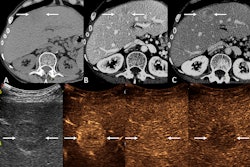
 Contrast-enhanced ultrasound (CEUS) Vesical Imaging-Reporting and Data System (VI-RADS) category 3 findings in a 40-year-old male with non-muscle-invasive high-grade urothelial carcinoma. (A) Grayscale US image obtained using a convex array probe (5C1) through a transabdominal approach showed a 35-mm lesion at the bladder posterior wall. (B) Color Doppler US image showed inner vascularity within the lesion. (C) Contrast-enhanced US image showed well-defined continuous layer of hypo-enhanced muscularis propria (white arrows) without vascular stalk on contrast-enhanced US or early hyper-enhanced continuous inner layer. The observation was assigned to VI-RADS 3 based on contrast-enhanced US by the readers devising the CEUS VI-RADS. Image and caption courtesy of the RSNA.
The research is important as CEUS has the potential to depict MIBC faster and at a lower cost, according to Glen Morrell, MD, PhD, of the University of Utah in Salt Lake City, who wrote a commentary that accompanied the study.
"In settings where MRI is not available, ultrasound may be a valuable alternative," Morrell wrote. "However, CEUS does have some relative disadvantages, which could limit its use ... CEUS requires the selection of a single image plane before injection of contrast material. This introduces the risk of missing muscle-invasive tumor at CEUS by undersampling. Additionally, CEUS is more invasive than MRI, requiring the use of intracavitary transrectal or transvaginal US probes for optimal depiction of bladder tumors."
The complete study can be found here.