Publicly insured pregnant women are less likely to get 20-week ultrasounds, a phenomenon that drives disparities in prenatal diagnosis of congenital heart defects, according to research published September 23 in Prenatal Diagnosis.
“To equitably improve prenatal diagnosis rates of congenital heart defects, investigation and intervention should focus specifically on improving rates of receiving second-trimester ultrasound,” noted a team led by Joyce Woo, MD, from Ann & Robert H. Lurie Children’s Hospital of Chicago.
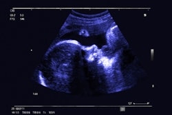
The 20-week ultrasound is an important exam in detecting birth defects, as it involves assessment of the major fetal organs. The exam is a routine part of prenatal care and is covered by Medicaid.
However, the researchers explained that pregnant women may still experience barriers to getting this ultrasound done, such as not being able to take time off work.
Woo and co-authors sought to better understand the mechanisms behind insurance-related disparities in prenatal diagnosis of a congenital heart defect via a study that study analyzed electronic health records of pregnant women whose infants received congenital heart defect surgery between 2019 and 2020 in Chicago. Their analysis included data from 496 pregnant women. Of these, 305 had a prenatal diagnosis of a fetal heart defect and 215 were publicly insured.
On bivariate regression analysis, the researchers found ties between public insurance and a 12.6% lower probability of prenatal diagnosis. On multivariable analysis, public insurance was associated with a 13.2% lower probability of having a second-trimester ultrasound performed. However, this was no longer associated with prenatal diagnosis after adjusting for women's receipt of a second-trimester ultrasound. The researchers noted that this suggests a possible mediation effect.
“Mediation analysis confirmed that second-trimester ultrasound receipt mediated 39% of the relationship between public insurance and prenatal diagnosis,” they wrote.
The study authors suggested that barriers to obtaining a fetal echocardiogram could be more readily overcome if patients with public insurance obtain their second-trimester ultrasound. This is due to suspected congenital heart defects on ultrasound being the most common indication for fetal echocardiograms.
"We found that pregnant individuals with public insurance are less likely to receive prenatal diagnosis of a congenital heart defect than individuals with private insurance, in part because those with public insurance are less likely to receive second-trimester ultrasound," Woo told AuntMinnie.com. "We have found in our previous research that this occurs for many reasons; some examples include patients not understanding how the second-trimester ultrasound differs from their other prenatal testing, the lack of appointment options, or lack of transportation. These are all areas that clinicians and clinician leaders can intervene upon to ensure that pregnant individuals have equal opportunity to receive this critical test. "
Still, the authors pointed out that there remain many knowledge gaps in understanding mechanisms behind socioeconomic-based disparities in prenatal diagnosis.
“Future investigation, policy, and public health efforts that focus on mitigating barriers to second-trimester ultrasound receipt could increase overall prenatal congenital heart defect diagnosis rates in an equitable manner,” they wrote.
The researchers are continuing to study this topic.
"Currently, our team is trying to better understand patient perceptions about second-trimester ultrasound, and regulation in ultrasound/echocardiogram quality," Woo said.
The full results can be found here.







![Examples of ultrasound findings and techniques. (A) Images in a 39-year-old male patient with a mass in the left thigh. The mass is heterogeneous on the B-mode US image (compared with the patient in D) and showed increased microvascularity (superb microvascular imaging [SMI]) and shear-wave elastography (SWE) values. Undifferentiated pleomorphic sarcoma was diagnosed at biopsy (with pleomorphic rhabdomyosarcoma in surgical specimen). (B) Images in an 18-year-old male patient with a mass in the left leg. The mass is hypoechoic on the B-mode image, with no other findings suggestive of malignancy. The lesion is in contact with the cortex of the tibia, which is slightly irregular. CT revealed a doubtful anteromedial tibial erosion. The microvascular study demonstrated high vascularization, suggestive of malignancy. Periosteal Ewing sarcoma was diagnosed with both histologic and immunohistochemical confirmation. (C) Images in a 69-year-old female patient with a lump growing on the outside of the left leg. Multiple SWE examinations were performed (please note the high values obtained in the measurements, whereas the color map highlights the stiffness relative to adjacent tissues). SMI showed areas of increased vascularization to target for sampling. Undifferentiated spindle cell sarcoma was diagnosed at biopsy, with residual leiomyosarcoma in the surgical specimen after neoadjuvant therapy. (D) Images in a 56-year-old female patient with a mass in the right thigh. The mass is heterogeneous at both B-mode ultrasound (similar to patient A) and MRI (coronal T2-weighted spectral attenuated inversion recovery [SPAIR]; T1-weighted pre-contrast and postcontrast imaging), which even shows uptake after the administration of paramagnetic contrast material, which is traditionally suggestive of malignancy. Low values at SMI and elastography are suggestive of benignity. Spindle cell lipoma was diagnosed at biopsy, with atypical spindle cell lipomatous tumor in the surgical specimen.](https://img.auntminnie.com/mindful/smg/workspaces/default/uploads/2026/08/images-radiol250278fig2.APCFLSvX6p.jpg?auto=format%2Ccompress&dpr=2&fit=crop&h=167&q=70&w=250)


