Mass enhancement on recombined images using contrast-enhanced mammography (CEM) is associated with malignancy, especially when linked to low-energy findings, a study published July 12 in Radiology found.
Researchers led by Geunwon Kim, PhD, from Beth Israel Deaconess Medical Center in Boston also found that enhancement findings on CEM images are independent of background parenchymal enhancement and breast density, meaning they can be used in diagnostic settings in all women.
"We found that enhancements without low energy correlates also turned out to be cancer, like on MRI. This highlights the utility of CEM, especially in settings where MRI is not as readily accessible," Kim told AuntMinnie.com.
CEM's use in recent years has become increasingly popular as researchers have touted improved cancer detection compared with conventional mammography. The study authors have also noted CEM's relatively low cost and better accessibility than supplemental MRI.
"For radiologists, CEM is an emerging tool that can help with detection and problem solving of complicated findings, especially in dense breasts," Kim said.
Like MRI though, CEM also shows enhancement for benign lesions, which may lead to unnecessary biopsies and false-positive findings. Little data exists on the associations between enhancement type and low-energy findings with malignancy, as well as BI-RADS descriptors.
Kim and colleagues wanted to find out the rates of malignant and benign enhancement findings on CEM with or without associated findings on low-energy images using BI-RADS.
They looked at retrospective data from 371 diagnostic CEM studies performed in 371 women with an average age of 54. Kim et al reported that for the total number, sensitivity was 95%, specificity was 67%, positive predictive value (PPV) was 55%, and negative predictive value of enhancement on CEM images was 97%.
Out of these, 190 CEM studies showed enhancement findings. The researchers found that enhancing lesions were more likely to be malignant when associated with low-energy findings (26% vs. 59%) (p = 0.001).
The team also found that among enhancement types, mass enhancement composed 71% (99 of 140) of all malignancies with a PPV of 63% when linked to low-energy findings. Foci, non-mass enhancement, and mass enhancement without low-energy findings had PPVs of 6%, 24%, and 38%, respectively. Non-mass enhancement was also more likely to be benign overall (PPV, 43%), but the likelihood of malignancy was higher (PPV = 82%) when linked to calcifications.
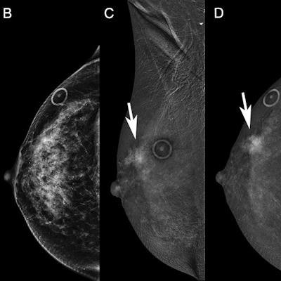
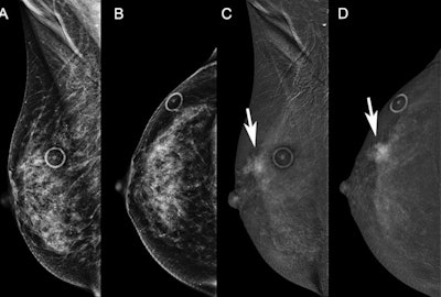
 Diagnostic contrast-enhanced mammograms (CEM) were performed on a 58-year-old woman recalled from screening for possible right breast distortion. Low-energy images on (A) mediolateral oblique and (B) craniocaudal views are unremarkable and the distortion did not persist. Recombined images show incidental mass enhancement (arrow) in the upper outer right breast on (C) mediolateral oblique and (D) craniocaudal views. These are examples of two-view recombined-only findings. Images courtesy of RSNA.
The study authors also wrote that using BI-RADS MRI lexicon to describe CEM recombined imaging findings showed "remarkably high" interobserver agreement (0.84) and can be used as a foundation for CEM BI-RADS. The team also reported that background parenchymal enhancement (p = 0.19) and breast density (p = 0.28) were not associated with enhancement type.
Kim told AuntMinnie.com that additional efforts to identify imaging features that could improve CEM positive predictive value and specificity are worthwhile. She added that this especially goes for targeting enhancing foci and low-energy asymmetries with associated enhancement.
"Dedicated analysis in a screening population would also be necessary, as CEM is increasingly being considered for this indication," the authors added.


