| FIGURE 1.1.3 Pedunculated advanced adenomas. 3D endoluminal CTC (A), coronal 2D CTC (B), and corresponding colonoscopy (C) images show a large pedunculated tubulovillous adenoma in the sigmoid colon. 3D endoluminal CTC image (D) from a second patie shows a 1.7-cm pedunculated polyp in the sigmoid colon, which is less conspicuous on the 2D CTC images (E, arrow) due to the similar appearance of hte sigmoid folds that are thickened by diverticular disease. The polyp was confirmed at same-day colonoscopy (F) and proved to be a tubulovillous adenoma. 3D endoluminal CTC (G) and double-contrast BE (H and I) images from three different patients show large pedunculated adenomas. |
Atlas of Gastrointestinal Imaging Figure 1.1.3 Pedunculated advanced adenomas
Latest in Home


Sponsored
Turning Innovation into Everyday Efficiency
April 22, 2026

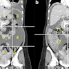


![A 53-year-old patient (patient number four) with a recurrent pituitary adenoma with extension of a cystic component of disease to the medial temporal lobe apparent on MRI (contoured in blue), and extension of disease to the left sphenoid bone and orbital apex apparent on [68Ga]Ga-DOTA-TATE (contoured in yellow).](https://img.auntminnie.com/mindful/smg/workspaces/default/uploads/2026/04/pituitary-tumor.QGsEnyB4bU.jpg?auto=format%2Ccompress&dpr=2&fit=crop&h=167&q=70&w=250)

